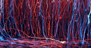
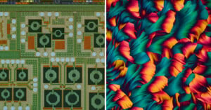
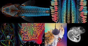
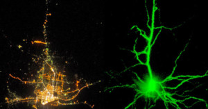
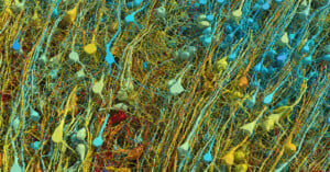
The Best Microscopic Life Science Photos of 2021
Olympus Image of the Year Award 2021 has announced the winners of its annual photography competition that recognizes the best worldwide talent in life science photos taken with a microscope.
Nikon has announced the winners of its 47th annual Small World Photomicrography Competition. This year’s first-place prize was awarded to Jason Kirk for his striking image of a southern live oak leaf’s trichomes, stomata, and vessels.
Olympus has announced the winners of its Global Image of the Year Life Science Light Microscopy Award, which is an annual competition that recognizes the best in life science photos taken with a microscope.
Nikon has unveiled the winners of the 2020 Nikon Small World Photomicrography Competition: our annual opportunity to witness the most beautiful images that straddle the intersection of stunning photography and groundbreaking science.
How do you go about capturing images of a coronavirus, which is too small to see with a standard light microscope? Here's a 5-minute video in which Vox explains how two electron microscopy techniques give us views of what the SARS-CoV-2 virus looks like.
A colorful photo of a tardigrade, popularly known as a "water bear," has won a top prize in the first-ever Olympus Global Image of the Year Award that was created to honor the best life science microscopy images.
In April of 2018, a team of scientists made an imaging breakthrough that allowed them to capture this incredible video of an immune cell moving through the tissues of a zebra fish's inner ear, picking up sugar particles as it goes along.
Nikon has announced the winning photos from the 2018 Nikon Small World Photomicrography Competition, the 44th annual contest celebrating "excellence in photography through the light microscope."
Photographer Linden Gledhill is a photographer who uses his background in biochemistry to capture gorgeous abstract images of the world at a microscopic level. His images are of various substances and chemical reactions, captured with high-tech microscope cameras that create focused stacked photos of extremely high resolutions.
When you look at the photographs in her series Into the Umbra, photographer Julia Bennett wants you to think you're looking at outer space. And then, just as your mind is struggling to expand to encompass the far reaches of the solar system where the image was captured, that's when she wants you to realize that you're looking at something you could find in any old liter of Sea Water.
Her images weren't captured with a telescope peering into the heavens, but a microscope that peers into the micro worlds inside droplets of seawater.
There is beauty in imperfection. In fact, imperfection might be considered the subject within a subject that photomicrographer Danny Sanchez tirelessly seeks out to create his stunning photography.
Sanchez's main subjects are gemstones, but the colorful, alien 'landscapes' he captures are made up of imperfections called 'inclusions' that actually make a gem less valuable. You might say that one gem merchant's trash is a gem photographer's treasure.
No, the title of this post wasn't written by some sort of broken record robot. It is in fact an accurate description of the GIF below, which was created from photographs taken with a Scanning Electron Microscope.
The intersection of Science, Technology and Art, at least according to renowned filmmaker and time-lapse photographer Louie Schwartzberg, is curiosity and wonder. And in the TED talk above, he makes the case for how few things pique that curiosity and inspire that wonder like the "hidden miracles of the natural world" that time-lapse, slow motion and microscopic imagery reveal.
It's always nice when we stumble across a copyright case that doesn't lead to wringing of hands and gnashing of teeth, rare as that might be. So when we ran across the news that a photographer pulled in $1.6 million in a copyright lawsuit, we just had to share it.
Science nerds and photographers can join hands today and stare in awe at what a team of researchers at the Department of Energy’s Lawrence Berkeley National Laboratory managed to do. Entirely by accident, these scientists have managed to take the first ever high-res images of carbon atoms in the process of forming chemical bonds.
David Scharf is a basement pioneer in the art of making some of the world's smallest things appear huge.
In the side-by-side images above, the photo on the left shows a city as seen by astronauts on the International Space Station, and then photo on the right shows a photo of a neuron imaged with fluorescence microscopy. One is massive and seen from a grand scale, while the other is microscopic and cannot be seen by the human eye, yet they look strangely similar in their structure.
Infinity Imagined has a gallery of these comparisons of cities and neurons, showing the strange and striking similarities between the two.
Gigapixel images are usually used to capture tiny details in expansive scenes, but scientists in the Netherlands recently created one that shows microscopic details in a tiny subject. Using a technique called virtual nanoscopy (a new relative of microscopy?), the researchers created a massive 281-gigapixel image of a 1.5-millimeter-long zebrafish embryo.
“Science can be beautiful. Art can be scientific.” This latest episode of the PBS series …
Photographer Clemens Wirth wanted to dive into microscopy, so he attached his Canon …