Neuron Formation Timelapse Wins Nikon Small World in Motion
Nikon Instruments Inc. has revealed the winners of the popular annual Nikon Small World in Motion Competition. This year’s awarded videos showcase the best in microscope videography, shedding light on the tiny unseen world.
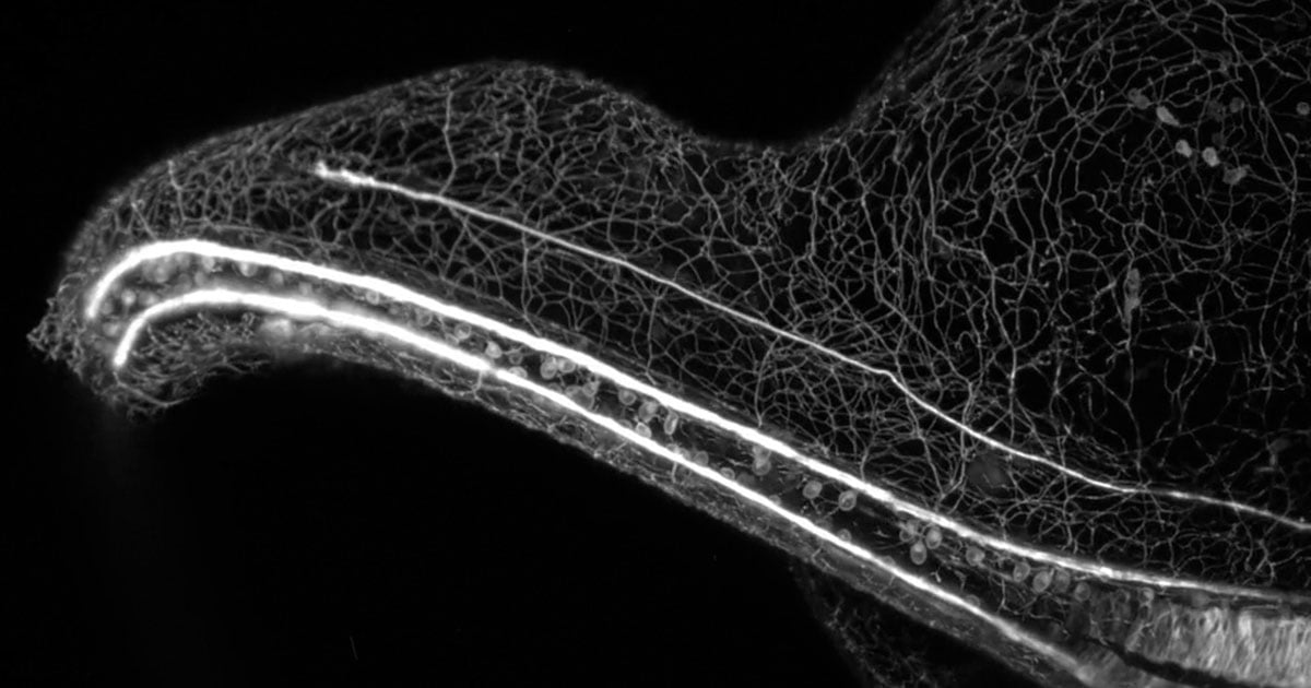
Dr. Alexandre Dumoulin has won first prize for his 48-hour timelapse video of developing neurons in a chick embryo. The neurons are connecting to the opposite side of the embryo’s central nervous system, and like they are in humans, the neurons are responsible for transmitting information throughout the body.
1st Place — Dr. Alexandre Dumoulin, University of Zurich, Department of Molecular Life Sciences, Zurich Switzerland. 48-hour timelapse of developing neurons connecting the opposite side of the central nervous system in a chick embryo. Confocal, 40x (objective lens magnification). | Nikon Small World in Motion Competition
Neurons are connected via long extensions known as axons, and axons travel throughout the nervous system before they form synapses. Dumoulin’s stunning video, developed at the University of Zurich, shows axons across the midline, or the boundary between the two hemispheres of the central nervous system.
For animals with neurological disorders, the movement of axons is impaired. There is even evidence that axon disorders, including abnormalities in axon guidance, may impact other conditions, including schizophrenia.
“My research focuses on investigating the developmental processes of neurons in chick and mouse embryos,” explains Dumoulin. “By studying these organisms, I aim to enhance our comprehension of how the nervous system functions and identify potential factors contributing to neurodevelopmental disorders.”
“The nervous system is an extremely complex and intricate system composed of a myriad of units that are connected to one another. In this video, we see single units and how they behave,” adds Dumoulin.
To capture the stunning video, Dr. Dumoulin utilized a new imaging method to visualize the live transfer of information from cells. “The biggest challenge was to discover a feasible method to access these neurons and capture images over an extended period of time,” he explains. “A combination of precise dissection skills and adapted microscopy techniques proved to be the key.”
The Nikon Small World in Motion Video Contest allows scientists like Dr. Dumoulin to share his research efforts and passion for microscopy. “I wanted to share these mesmerizing developing neurons with the public. To me, that’s the essence of this competition, highlighting the beauty of nature through the lens of scientific research,” he says.
“For nearly half a century, we’ve received awe-inspiring entries that are not only visually stunning but scientifically groundbreaking,” says Eric Flem, Senior Manager, CRM and Communications at Nikon Instruments. “This year’s winning video is no different; while beautiful, Dumoulin’s entry can carry significant meaning for the advancement of potential treatments for neurodevelopmental diseases.”
Stunning Videos Round Out an Impressive Top Five
Alongside Dumoulin’s prize-winning entry, the competition also names second, third, fourth, and fifth place finishers, alongside a large selection of honorable mentions.
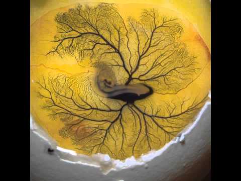
2nd Place — Fabian J. Weston, Protist Lab Films, Pennant Hills, New South Wales, Australia. Blood flow in the tail fin of a small fish. Differential Interference Contrast (DIC). 10x (Objective Lens Magnification). | Nikon Small World in Motion Competition
Fabian J. Weston of Protist Lab Films has earned second place with his video of blood flow within the tail fin of a small fish. Weston administered filtered sample water with oxygen throughout the filming process.
In third place is Nell Saunders with the Institut Pasteur for her video of human cells fusing and dying after being infected by SARS-CoV-2.
3rd Place — Nell Saunders, Institut Pasteur, Department of Virology, Paris, France. Human cells fuse and die upon infection by SARS-CoV-2. Holotomography, 60x (objective lens magnification). | Nikon Small World in Motion Competition
Rounding out the top five is Benedikt Pleyer in fourth place with his footage of pond creatures and Dr. Michael Weber in fifth place, whose award-winning video shows the beating heart of a five-day-old zebra fish.
4th Place — Benedikt Pleyer, Kirchberg, Bavaria, Germany. Pond creaturs (in order of appearance): Hydra, Volvox, Daphnia, Spirostomum, Synura, Hydra. Darkfield, 2.5x-3.5x (objective lens magnifications). | Nikon Small World in Motion Competition
5th Place — Dr. Michael Weber, Georg-August-University Göttingen, Multiscale Biology, Göttingen, Niedersachsen, Germany. The beating heart of a five-day-old zebra fish. Light seet, 20x (objective lens magnification). | Nikon Small World in Motion Competition
All winning videos in the 2023 Nikon Small World in Motion competition, plus winners from prior years, are available on the Nikon Small World website. The winners of this year’s Small World Photomicrography Competition should be announced soon.
Credits: Videos courtesy of Nikon Instruments Inc.