Winning Photos from the 2018 Nikon Small World Competition
Nikon has announced the winning photos from the 2018 Nikon Small World Photomicrography Competition, the 44th annual contest celebrating “excellence in photography through the light microscope.”
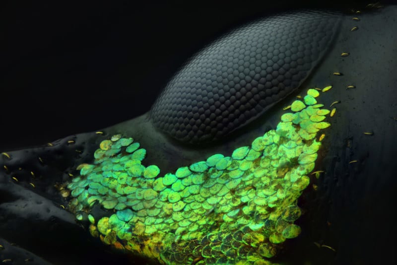
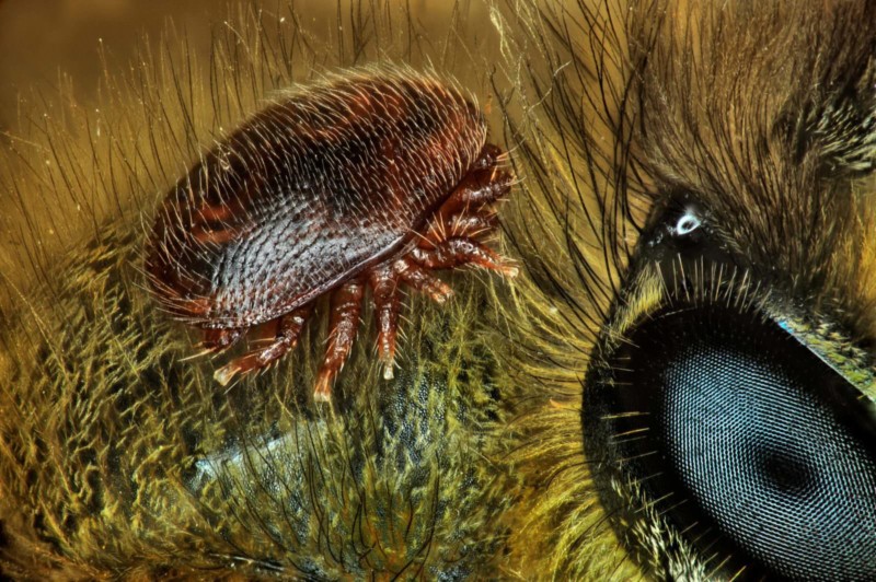
Al Habshi created the photo using reflected light and by stacking 128 micrographs into a single photo.
“The main challenge was to show the black body against the black background without overexposing the skin and scales,” the photographer says. “Because of the variety of coloring and the lines that display in the eyes of insects, I feel like I’m photographing a collection of jewelry. Not all people appreciate small species, particularly insects.
“Through photomicrography we can find a whole new, beautiful world which hasn’t been seen before. It’s like discovering what lies under the ocean’s surface.”
Here is a selection of other winning photos from this year’s competition:
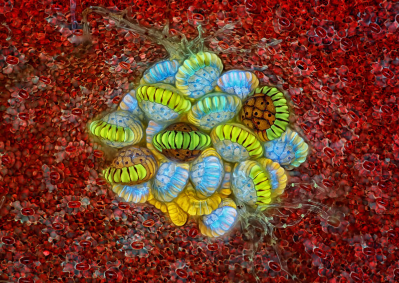
Spore Structures
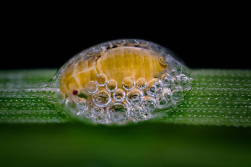
Bug in Bubble House
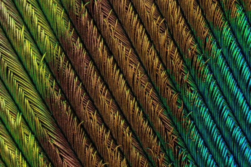
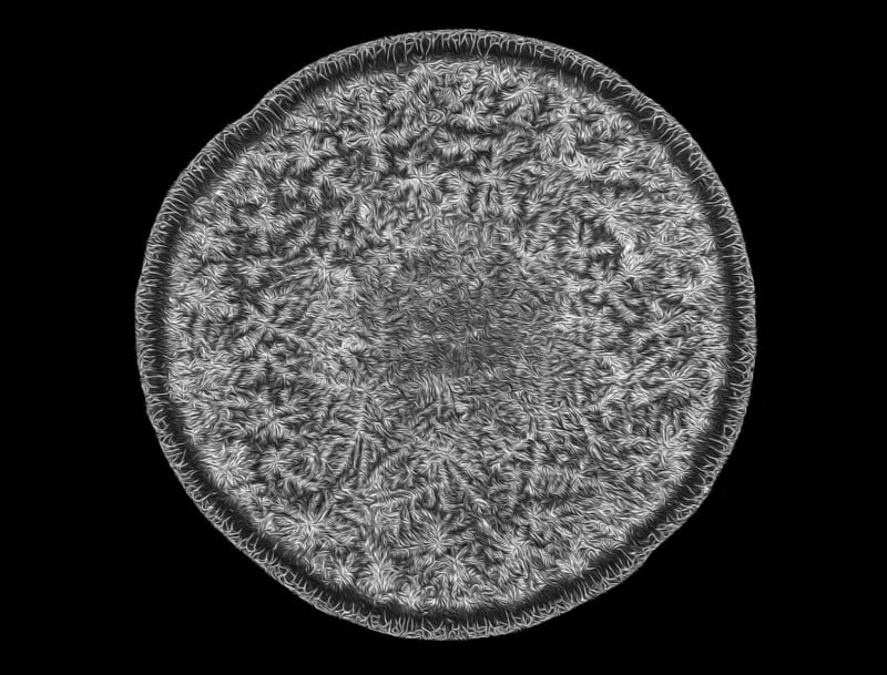
Peacock Feather
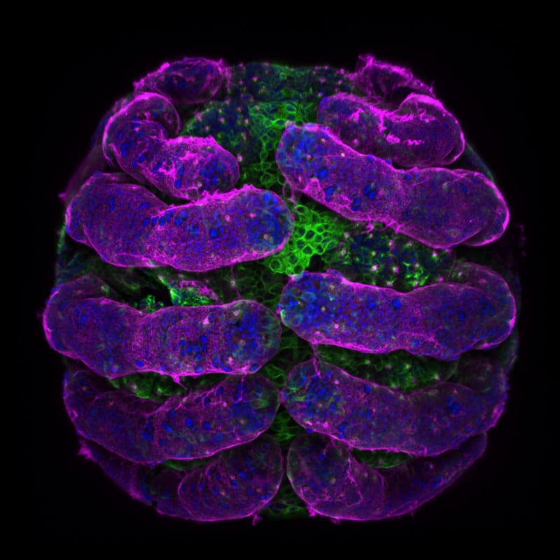
Spider Embryo
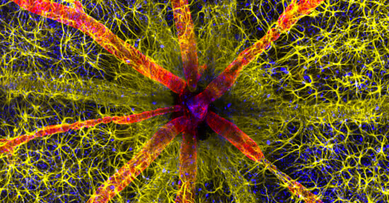
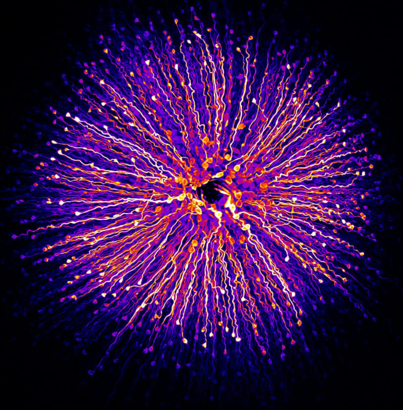
Retina
Tear Drop
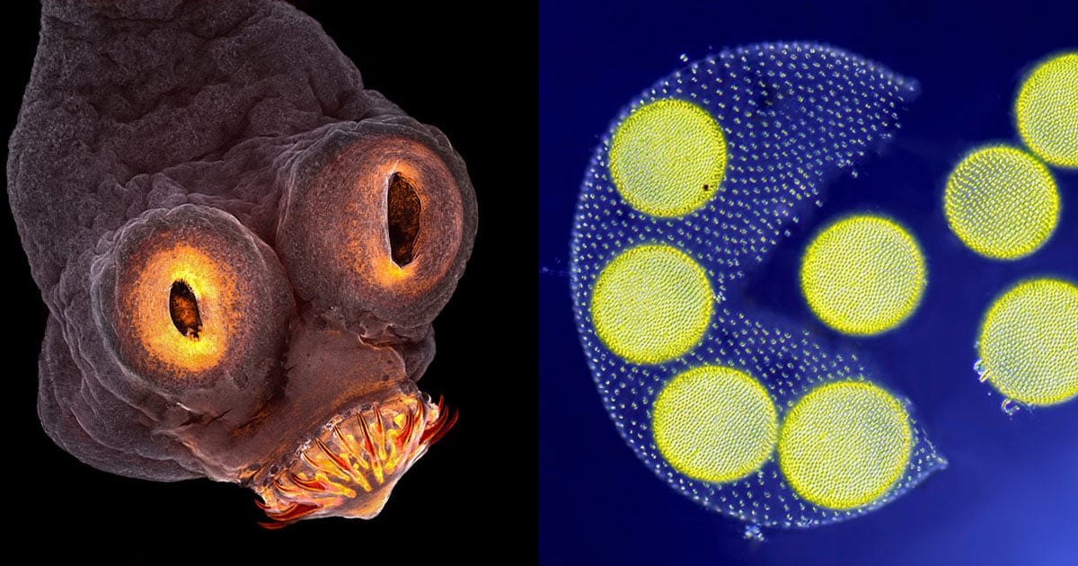
Weevil Portrait

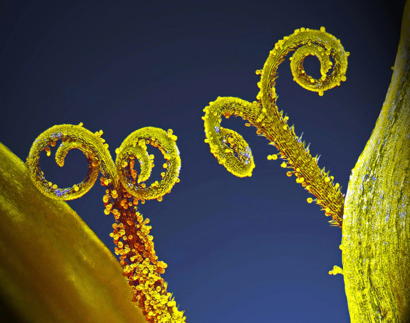
Stalks and Pollen
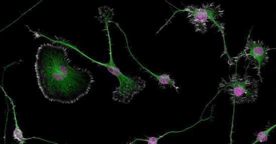
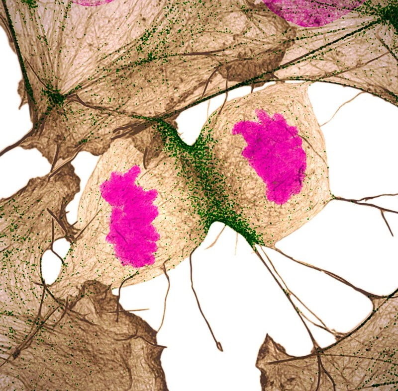
Human Cell Division
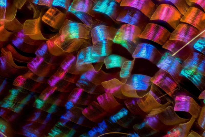
Butterfly Wing Scales
A Mite on a Bee
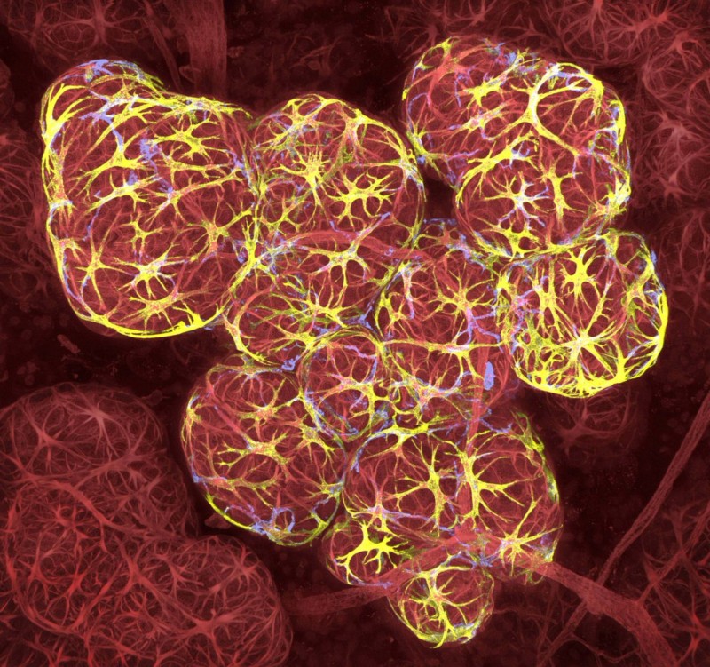
Breast Tissue in Lactation
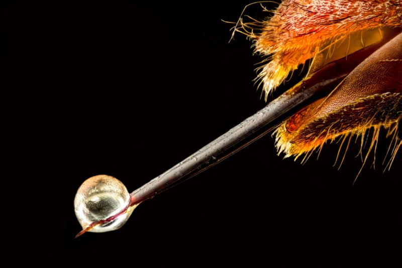
Hornet Venom
You can find the complete gallery of winning photos on the Nikon Small World Competition website.