

Canon Technology Used to Perform World’s First Coral ‘Pregnancy Test’
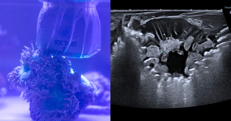
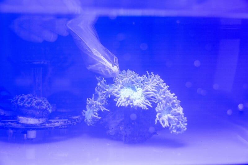
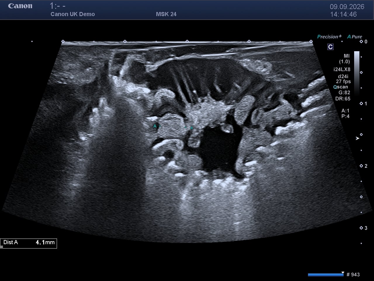
Coral Spawning International (CSI), in partnership with Canon Medical Systems UK and Canon EMEA, has successfully conducted the world’s first ultrasound scan of living coral to identify developing eggs.
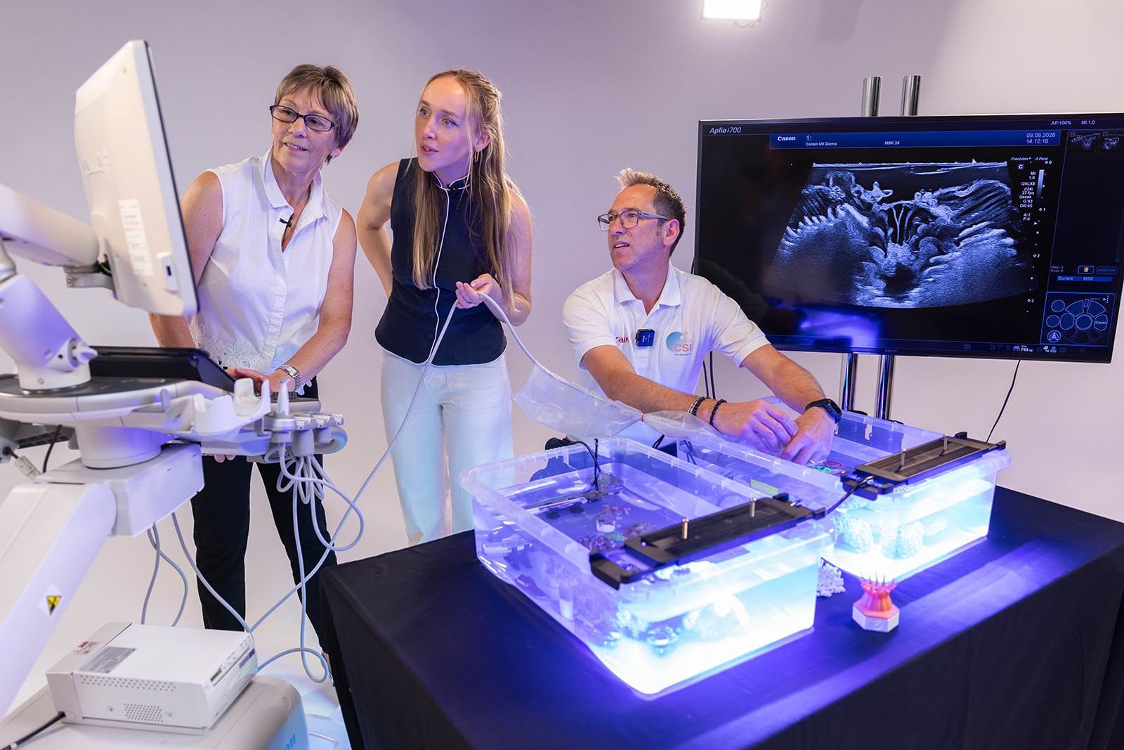
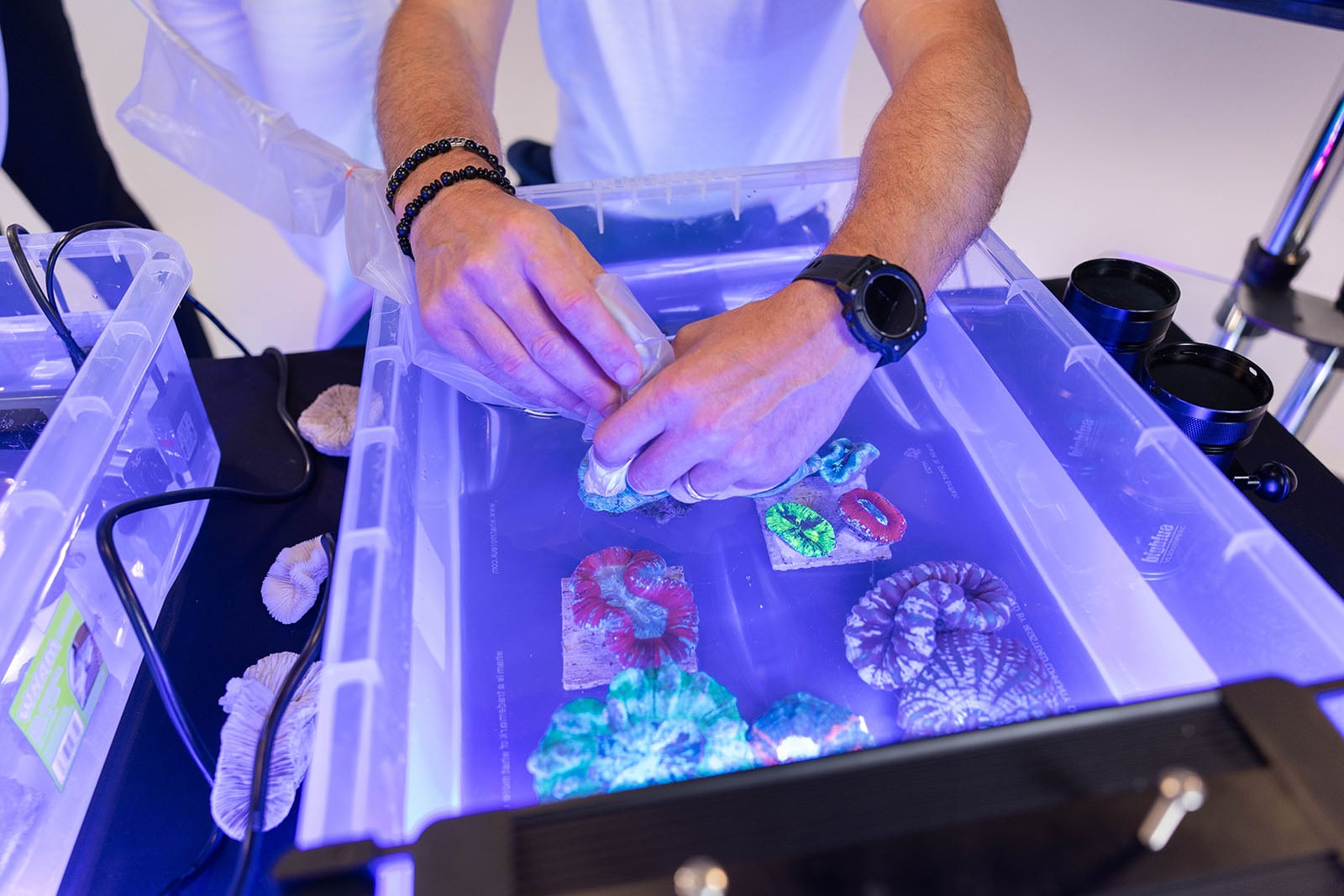
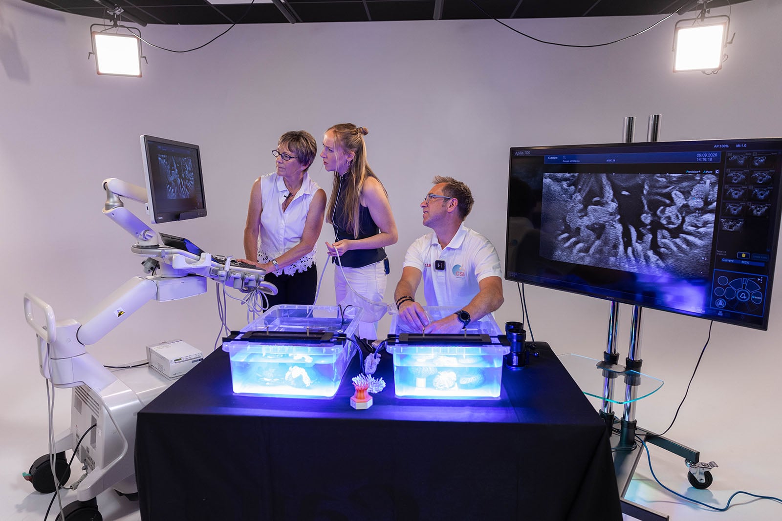
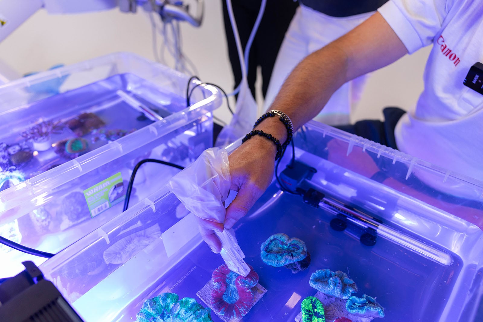
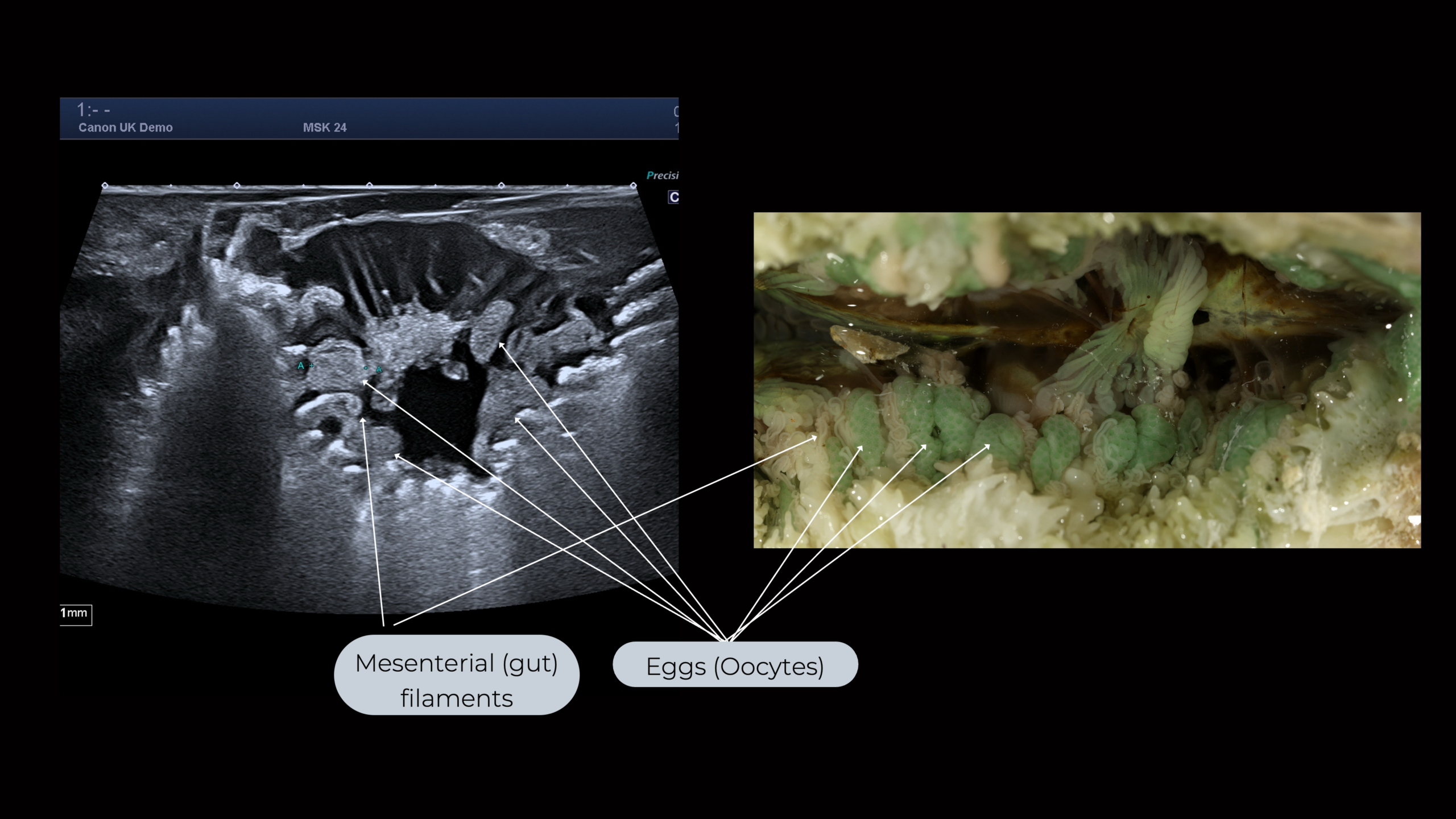
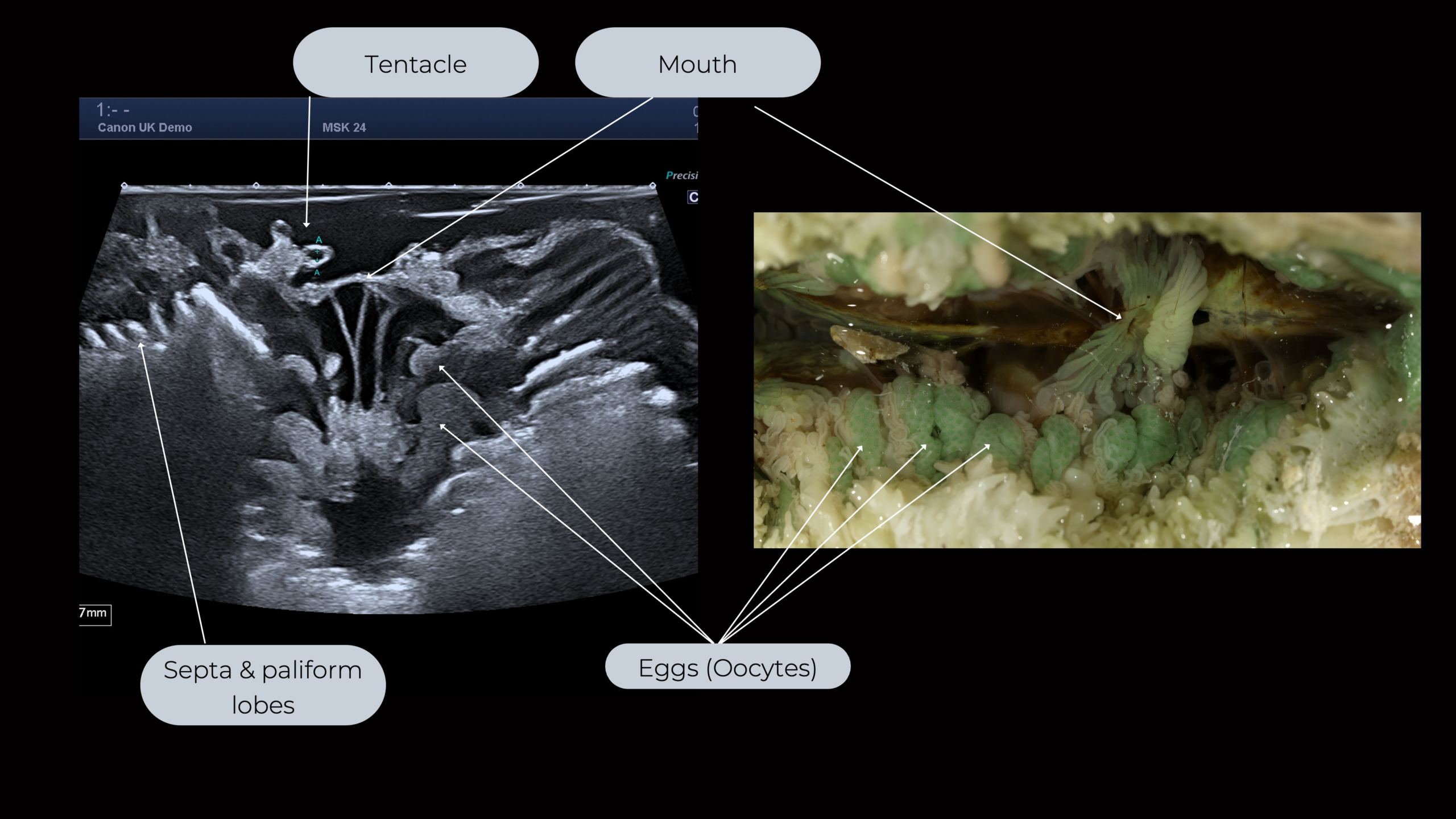
At a time when coral conservation is of paramount importance, this is the first non-invasive ultrasound scan on living, submerged corals that has been completed. It was conducted with the Aplio i700, an advanced Canon ultrasound platform. Scientists explain that accurately detecting egg development in living colonies allows researchers to better predict spawning windows, accelerating lab-based propagation, selective breeding, and reef restoration efforts.
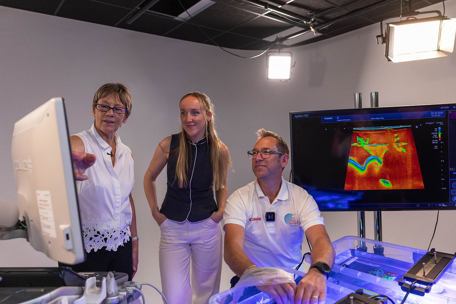
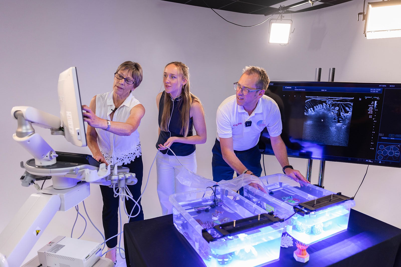
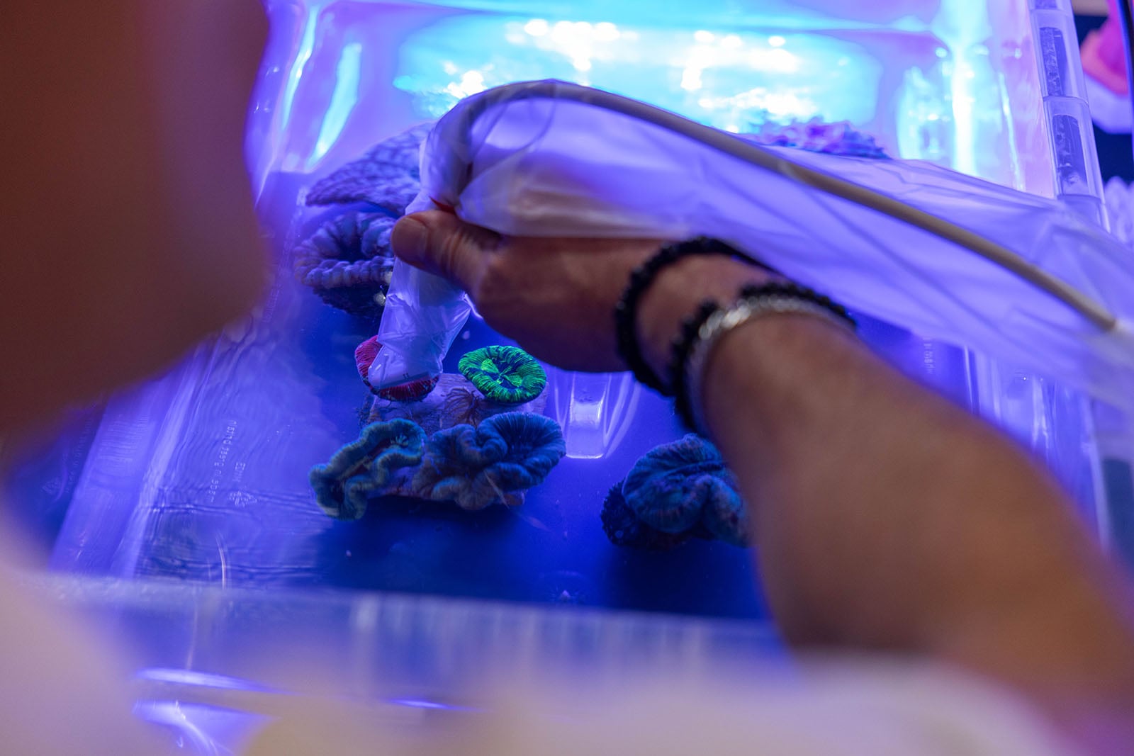
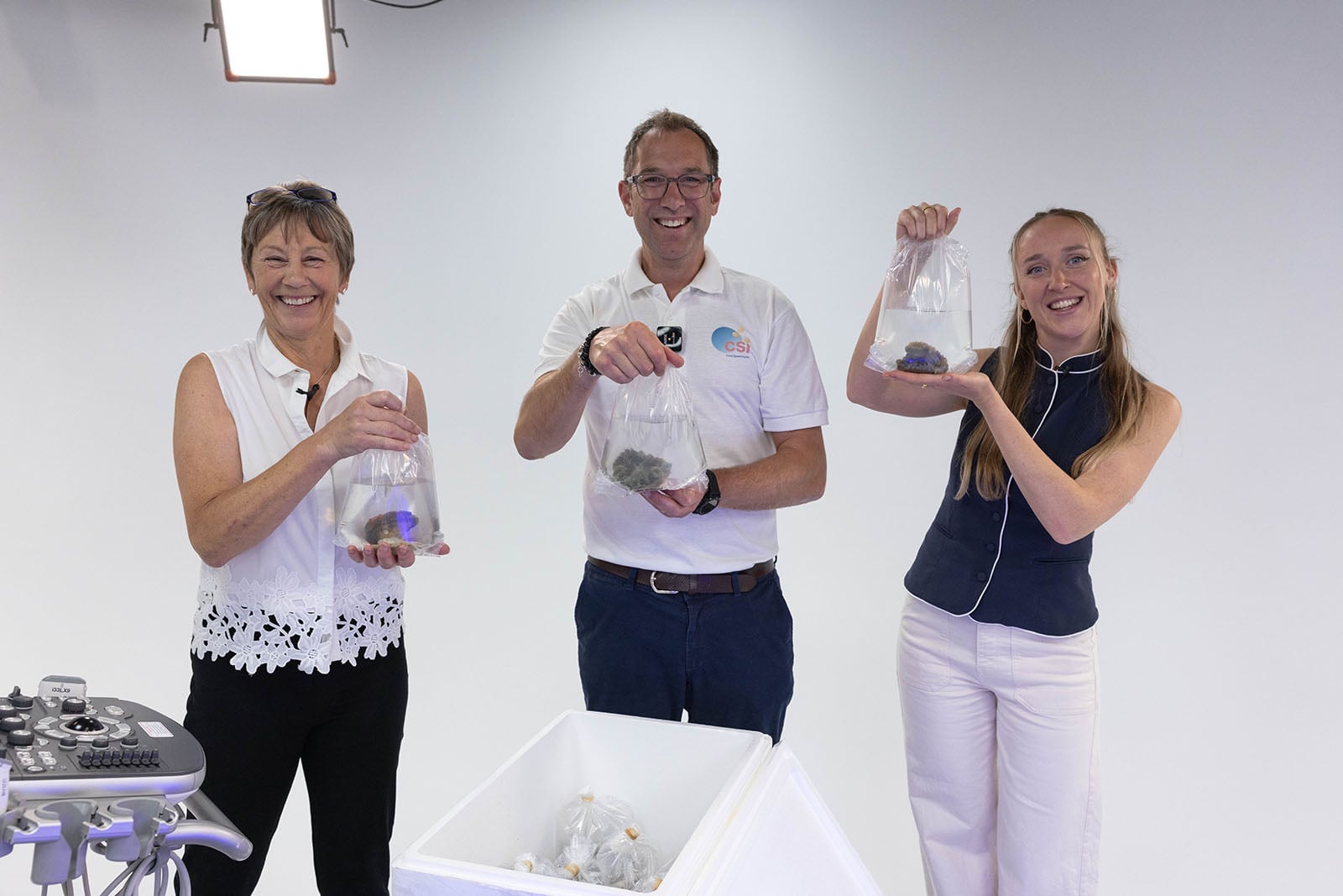
“Corals still hold so many secrets, particularly when it comes to the complex processes that drive their reproduction. Being able to look inside a living coral and see these processes unfolding in such remarkable detail is an extraordinary moment. This experiment has given us a new perspective on these animals and, as we continue monitoring them through spawning, we hope to build an increasingly detailed picture of how different coral species reproduce,” Dr. Jamie Craggs, Co-Founder of Coral Spawning International, says.
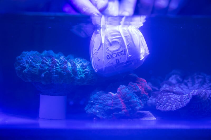
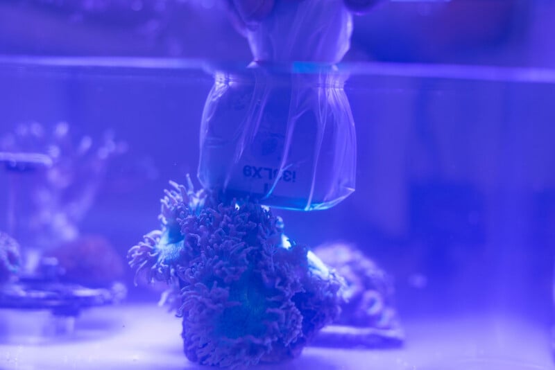
“This experiment showcased just how powerful ultrasound technology can be. Moreover, that the experiment was successful opens opportunities in fields such as minimally invasive surgery and diagnosis of dermal conditions,” Sally Davey, Clinical Product Specialist, Canon Medical Systems UK, adds.
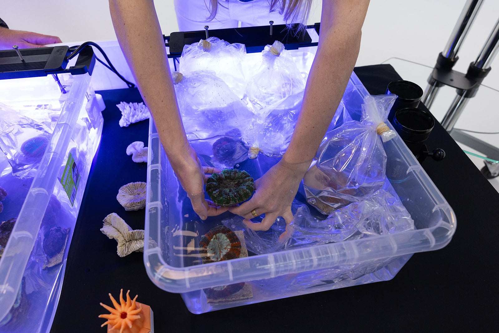
Using Canon’s Aplio i700 hospital ultrasound system with a mix of high and ultra-high frequency probes, researchers successfully imaged internal egg structures in six large polyp stony (LPS) coral species underwater, which eliminates the traditional need to cut open or sacrifice living corals to check their reproductive state. The scan findings were verified on Trachyphyllia geoffroyi coral by cross-referencing the ultrasound images with macro photography.
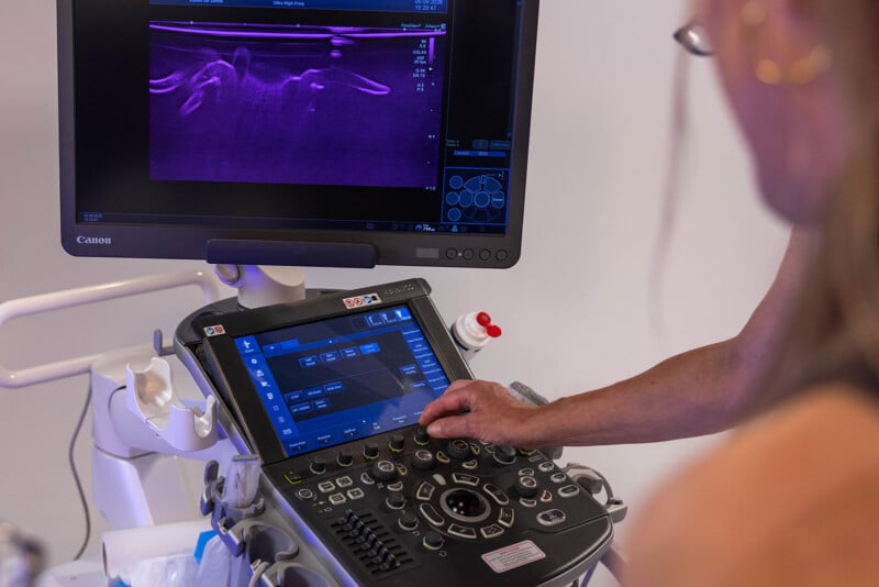
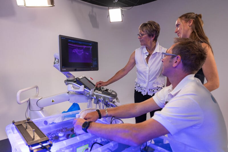
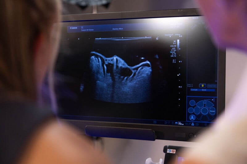
While this achievement is impressive on its own, Canon says that the success of high-resolution underwater ultrasound provides valuable insights for human medical fields, including minimally invasive surgery and dermal diagnostics.
“This experiment demonstrates that imaging innovation has the power to show us what was previously unseen, creating deeper understanding of the world around us. By unlocking this knowledge, we hold a fundamental belief that imaging has the power to transform the world,” Peter Bragg, Sustainability and Government Affairs Director at Canon EMEA, explains.
Canon adds that the corals used in this experiment will continue to be monitored for six months, with recordings taken when gametes (mature reproductive cells) are released. This will allow CSI to better understand the coral reproductive cycle, helping identify spawning windows more accurately and support future coral conservation and restoration efforts.
Image creditsCanon EMEA