
A New Super-Resolution Microscopy Technique Lets Scientists Study Cells In Real Time
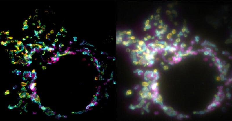
A new fluorescence microscopy technique promises to create super-resolution images from a single exposure and give scientists a remarkable look inside cells.
The technique, reported by Phys, is called SPIFFI. This stands for spatial polarization-induced fluorescence fluctuation imaging, as researchers Wei Guo, Lely Feletti, and Aleksandra Radenovic explain.
The research paper about SPIFFI was recently published in Nature and goes into extensive detail about the technique. While the nitty-gritty science is far beyond the scope of this article, the primary takeaway is that SPIFFI relies upon the polarization of fluorescent light to overcome challenges associated with traditional super-resolution microscopy.
Super-resolution microscopy has been transformative in cell biology, enabling scientists to investigate cellular structures that are far too small to see with conventional light microscopes. However, traditional methods often rely on multi-frame capture, like hundreds or even thousands of individual images. This makes imaging living cells very challenging, if not impossible. Living cellular structures are always moving.
Radenovic, who works in the Laboratory of Nanoscale Biology (LBEN) in EPFL’s School of Engineering explains that SPIFFI “can capture fast-moving processes within cells, while enabling high-throughput, multi-dimensional imaging beyond the limits of conventional microscopes.”
“Essentially, previous approaches used temporal information to resolve spatial resolution, but this doesn’t work very well on living cells,” adds first author and LBEN PhD student Wei Guo.
SPIFFI relies on the fact that fluorescent molecules emit polarized light, meaning light waves emitted by a molecule “oscillate preferentially in certain directions, depending on how the molecule is oriented.”
SPIFFI “splits this fluorescent light into four polarization-sensitive channels, and compares the resulting images to recover previously hidden structural details.”
The researchers demonstrated through experiments that SPIFFI can improve image resolution by a factor of two in a single image, rather than hundreds or thousands of them. It can resolve structures about 160-170 nanometers in size and has been used to sharply image events like cellular splitting and fusion.
“With previous techniques, taking many images would only result in one super-resolved frame. With SPIFFI, every frame is super-resolved, meaning we can now produce super-resolution videos of live cells,” Guo continues. “We also seamlessly integrated SPIFFI images with existing fluctuation-based methods for post-processing, achieving resolutions of about 80 nanometers.”
Promisingly, SPIFFI’s optical hardware can also be implemented in existing fluorescence microscopes, making it far more practical than a full-fledged, dedicated setup. The researchers are now working to make SPIFFI more compact, which will further improve its usability.
Image creditsEPFL
Affiliate Disclosure PetaPixel articles may include affiliate links; we may earn a commission if you buy through one.