How X-Ray Photographer Jim Wehtje Sees Beneath the Surface of Seashells
Jim Wehtje is an American photographer and radiographic artist known for using X-rays to reveal the hidden internal structures of natural objects, particularly plants, flowers, and seashells.
Raised in Massachusetts, Wehtje studied natural sciences before moving to Rochester, New York, in 1994 to study biomedical photography at the Rochester Institute of Technology (RIT). At the recommendation of Professor Michael Peres (now retired), he was introduced to research equipment at a local hospital. This allowed him to start his creative X-ray imaging career.
Wehtje began creating X-ray photographs in 1996. His background in both science and photography led him to explore radiography not simply as a diagnostic tool, but as a photographic medium. He became an early commercial practitioner of non-medical radiography, marketing X-ray images through Photodisc, later part of Getty Images, and producing work for commercial and editorial clients. In 1998, his work appeared on the cover and served as the theme of the Photodisc.com/2 catalog, which was still printed and mailed to customers at the time. The exposure from the catalog helped bring Jim’s work to a much wider audience and ultimately led to his X-ray images of starfish being featured on the packaging for Adobe® GoLive CS2 in 2004.
Jim still uses Photoshop, but as a protest against constant upgrades, he runs version CS6 (2012), which is on a perpetual license — with no monthly charges. This forces him to keep his main workstation Mac on System 10.8.5 to run it.
“I changed the hard drive once and hope it lasts,” says Wehtje.
Much of Jim’s work concentrates on botanical specimens and seashells. Because delicate plants absorb relatively little X-ray radiation, he uses specialized equipment capable of producing low-energy X-rays to obtain sufficient detail and contrast. For subjects with a wide range of densities, he may make multiple radiographic exposures and digitally combine them, somewhat analogous to HDR photography. He also digitally assembles individual specimens into larger compositions and sometimes adds color to the monochrome radiographs.
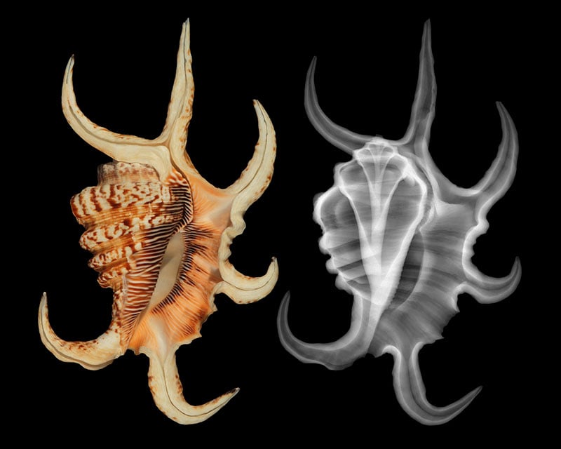
Wehtje describes radiography as an intersection of science and art. Rather than recording the surface appearance of a subject, his images reveal structures normally hidden from human vision — the veins of leaves, internal organization of flowers, and intricate chambers and spirals within shells. He continues to work from Lancaster, Massachusetts, and has been producing X-ray imagery for approximately three decades.
His recent work continues to receive attention. In August 2026, National Geographic featured his image of an arthritic spider conch (Harpago arthriticus), associated with an upcoming exhibition at the Bailey-Matthews National Shell Museum in Sanibel, Florida.
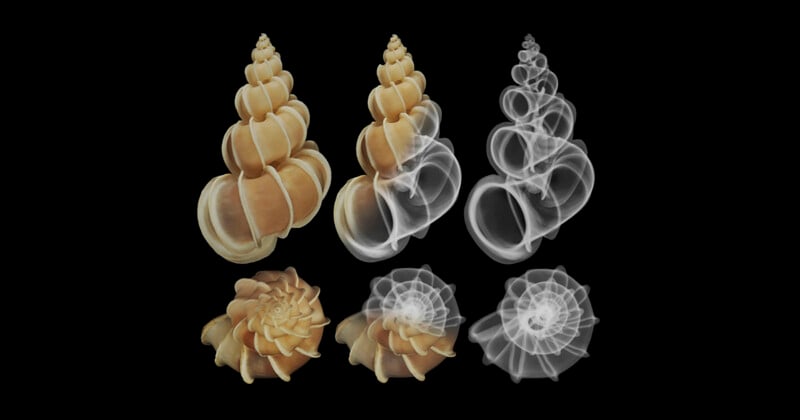
The images compare and often blend X-ray and traditional reflective-light images of each shell specimen. Radiography is, of course, a classic imaging medium for shells, revealing their intricate internal structures.
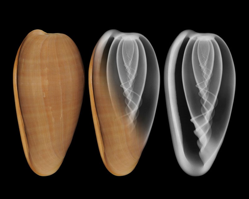
In January, Jim released Earth Shell Forms X-Rayed, which is currently self-published print-on-demand here. The book took over 14 months to create and is a romp through mollusk shells and beyond to plastic and glass enclosures. It “does not shy away from ecological, mythical, and philosophical implications, which are given from an outsider universalistic perspective,” says Jim.
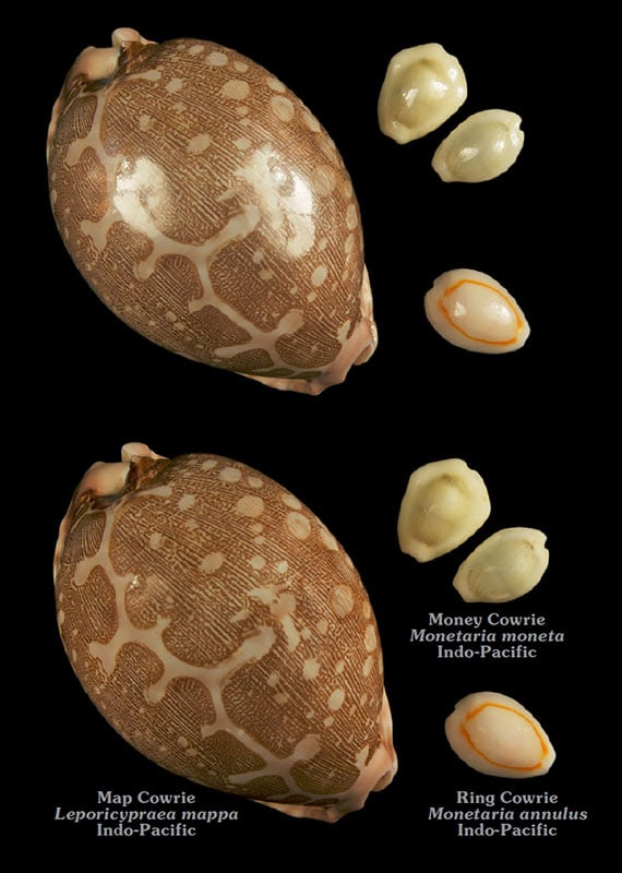
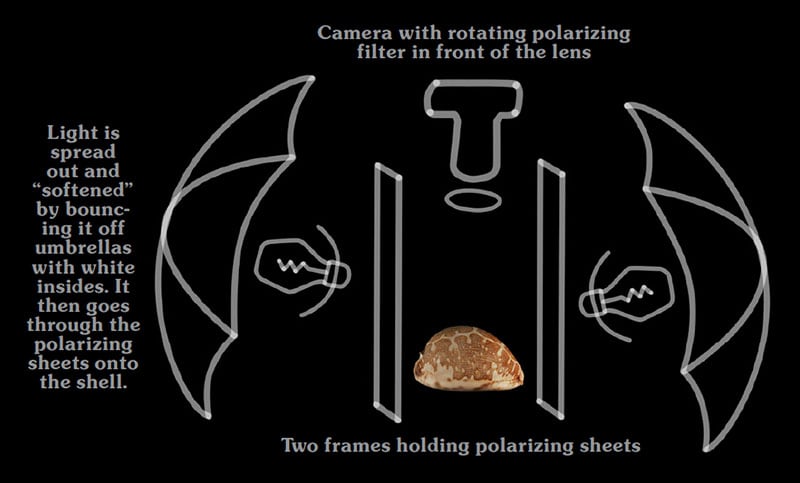
As an X-ray specialist, Wehtje thought he would never use his camera again. But for the book project, he dusted it off to make more of the color images to pair with the X-ray ones — rediscovering color. This work naturally led him to submit the imagery to the Bailey-Matthews Shell Museum. Technically, Wehtje found the round, shiny cowries the hardest shell family to photograph. He used a cross-polarized light setup to eliminate direct reflections.
Jim lives in Lancaster, Massachusetts, with an extensive garden that supplies him with a never-ending source of subjects and inspiration. His website is a rich portfolio of Wehtje’s prolific career, featuring X-ray photographs of diverse subjects, natural and human-made alike.
An exhibit of 23 X-ray photos of shells by Jim Wehtje opens October 10 at the Bailey-Matthews National Shell Museum & Aquarium in Sanibel, Florida.
Image creditsJim Wehtje
Affiliate Disclosure PetaPixel articles may include affiliate links; we may earn a commission if you buy through one.