17 Award-Winning Microscope Photos Reveal the World’s Hidden Wonders

A collage of three microscopic images: a colorful cell with hair-like projections, a glowing blue spherical cluster with radiating lines, and a textured orange and purple layered surface.

Evident Scientific, a scientific solutions and microscopic imaging company, has announced the winners of its sixth annual Image of the Year photo contest. The competition celebrates the world’s best scientific microscopic imaging, and the photos are as scientifically valuable as they are beautiful.

While traditional photographers may not know much about Evident, they all know the company it used to be. Olympus Corporation spun off its Scientific Solutions business a few years ago into a wholly owned subsidiary, Evident, as part of Olympus’ broader reshuffling. This same divestment process also led to the creation of OM Digital Solutions, makers of OM System cameras and lenses.

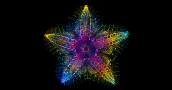
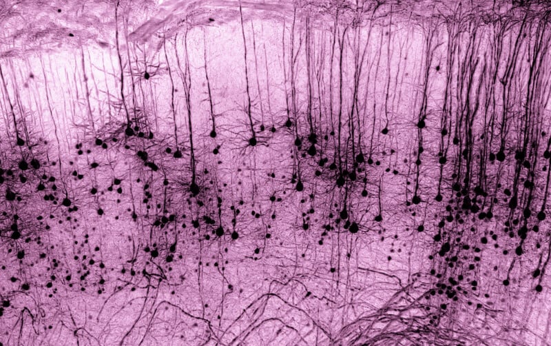
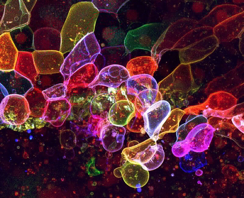
Evident’s latest Image of the Year competition attracted entries from 34 different countries, with Katie Holden from the United Kingdom taking top honors for her visually striking image, “Neuronal Cosmos.”

A bright, glowing blue sphere with radiating electric-like lines and wisps, set against a dark background, resembling a neural network or energy burst.
Global Winner, “Neuronal Cosmos,” by © Katie Holden | Evident Image of the Year 2026

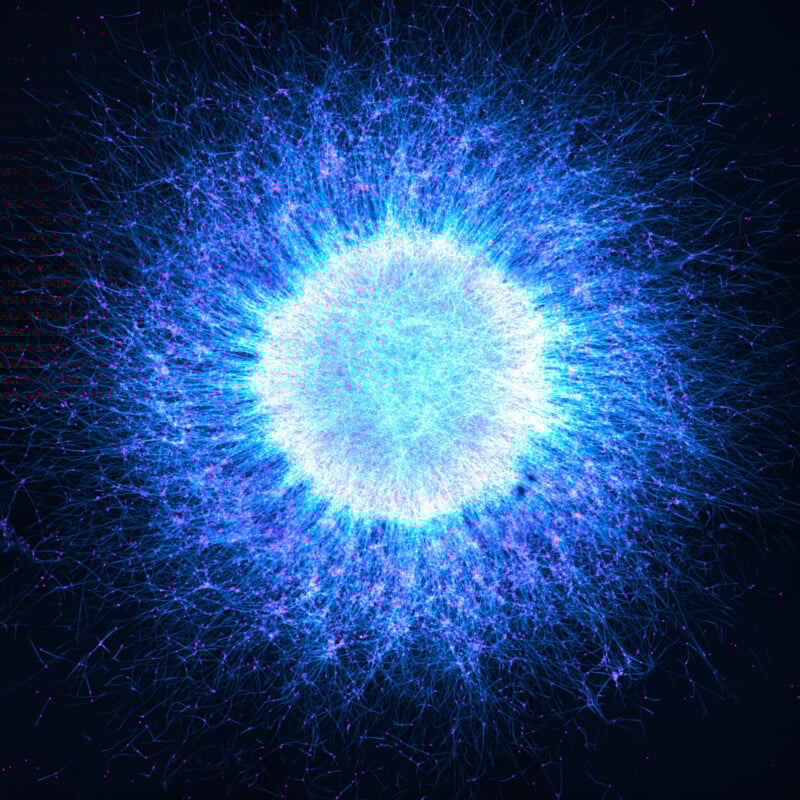
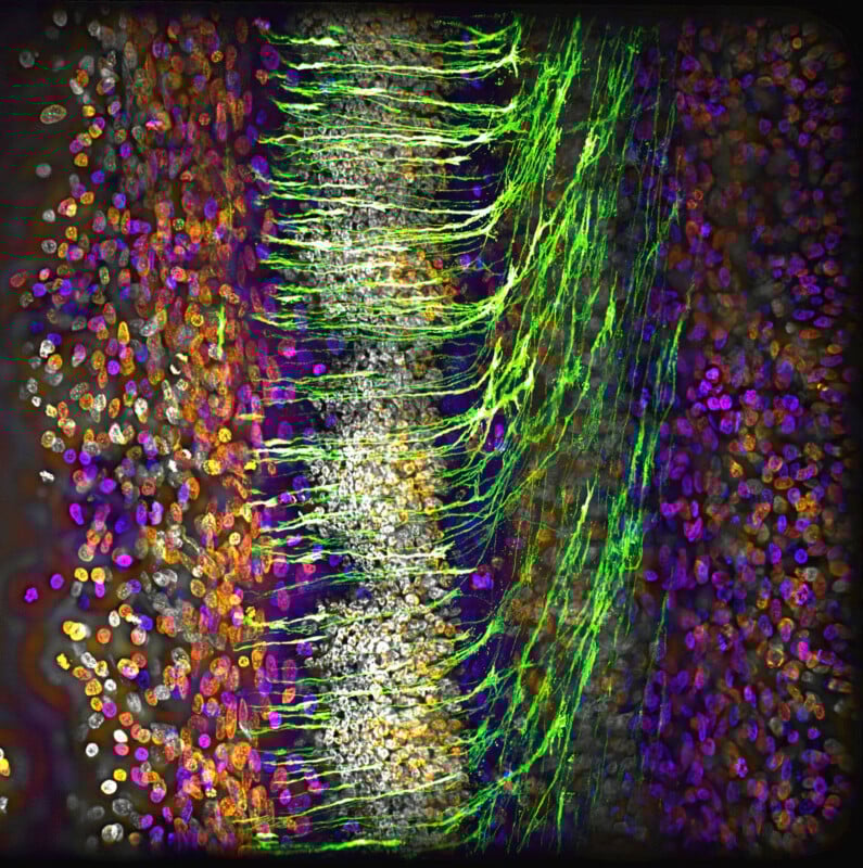
Holden’s photo shows induced pluripotent stem cell-derived neurospheres, Evident explains. These neurospheres comprise neuronal cells, which organize into “structures resembling the layered architecture of the human brain.”

Neurospheres are an important way for scientists to study brain cells, including researching how environmental and genetic factors influence neuronal development.

“Visually, the star-like pattern reflects intriguing parallels between astronomy and biology at hugely different scales,” Holden explains. She will receive an Evident SZX7 stereo microscope and a DP23 digital camera, or a set of X Line UPLXAPO objectives. The DP23 camera is a 6.4-megapixel CCD color camera that still carries Olympus branding, a testament to its heritage.

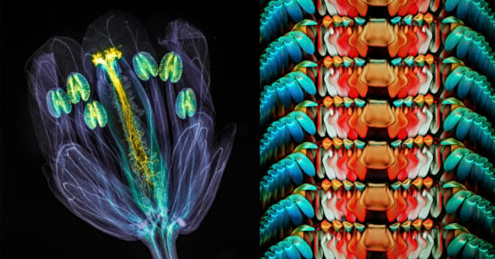
The competition also features other specific winners, including Muhammad Tahir Khan from Ireland, who won the Materials Science category for an image of lignin fiber that looks like glowing desert dunes. Khan receives an Evident SZ61 stereo microscope as a prize.

Orange and purple sand dunes form sweeping, wavy patterns, with vibrant ridges and soft gradients creating an abstract, surreal desert landscape under a colorful sky.
Materials Science Winner, © Muhammad Tahir Khan

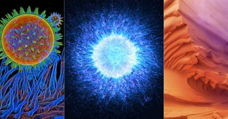
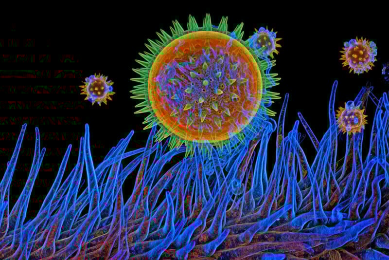
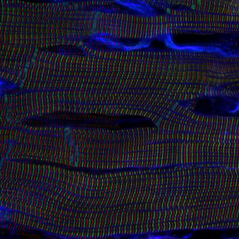
There are also three Regional Winners. Gerd Günther from Germany won the EMEA region for an image of a chicory stigma with pollen grains. American Igor Siwanowicz won the America category for a microscopic photo of mallow pollen. Kentaro Mochizuki of Japan took home the Asia Pacific regional prize for an incredible shot of sarcomere structures in a rat heart. Each regional winner will choose between a CX23 upright microscope or a SZ61 stereo microscope.

A close-up microscopic image of a pale, spiral-shaped plant stem covered in tiny hairs, surrounded by clusters of bright, blue, star-like pollen grains on a black background.
EMEA Regional Winner, © Gerd Günther
Colorful, computer-generated illustration of a virus particle attaching to hair-like structures on a cell surface, against a black background. The virus is shown in orange and green, while the cell surface is blue and purple.
Americas Regional Winner, © Igor Siwanowicz
Microscopic view of muscle tissue displaying parallel, wavy lines in red, green, and blue with irregular, dark horizontal gaps and blue highlights, illustrating the striated pattern of muscle fibers.
APAC Regional Winner, © Kentaro Mochizuki

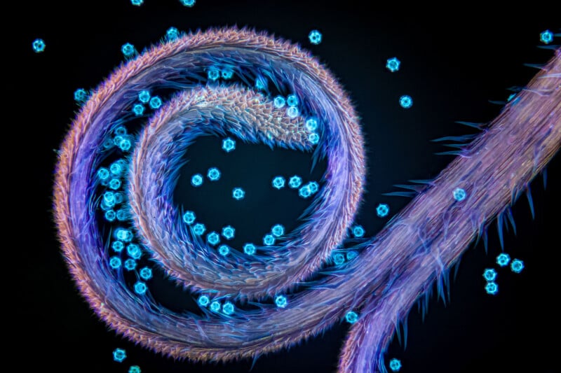
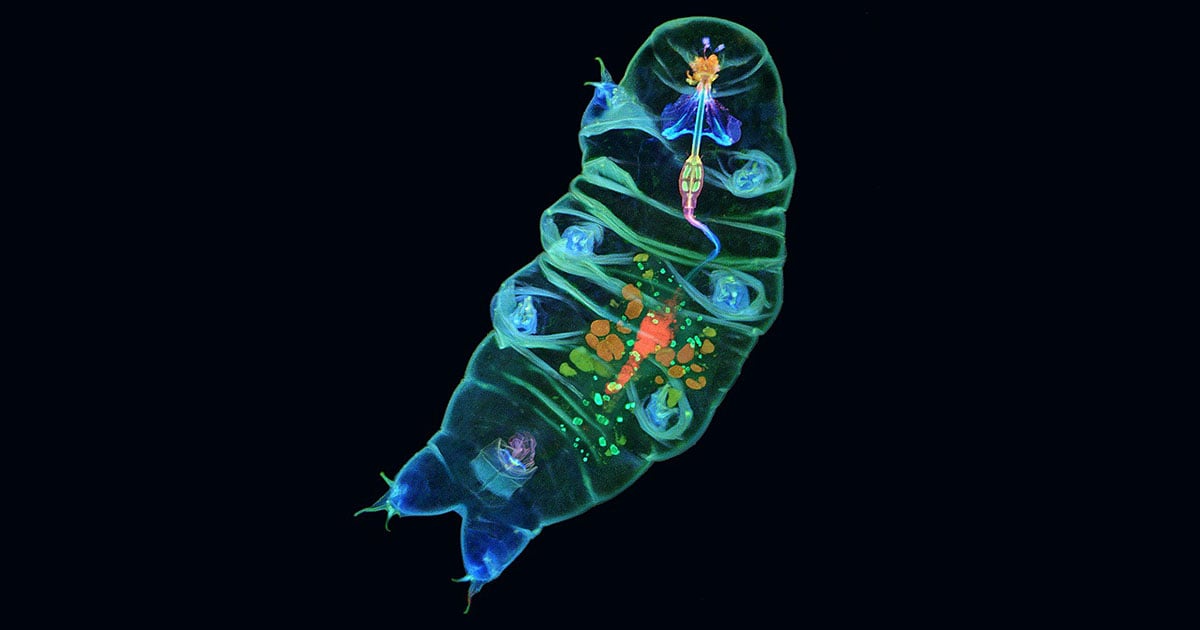
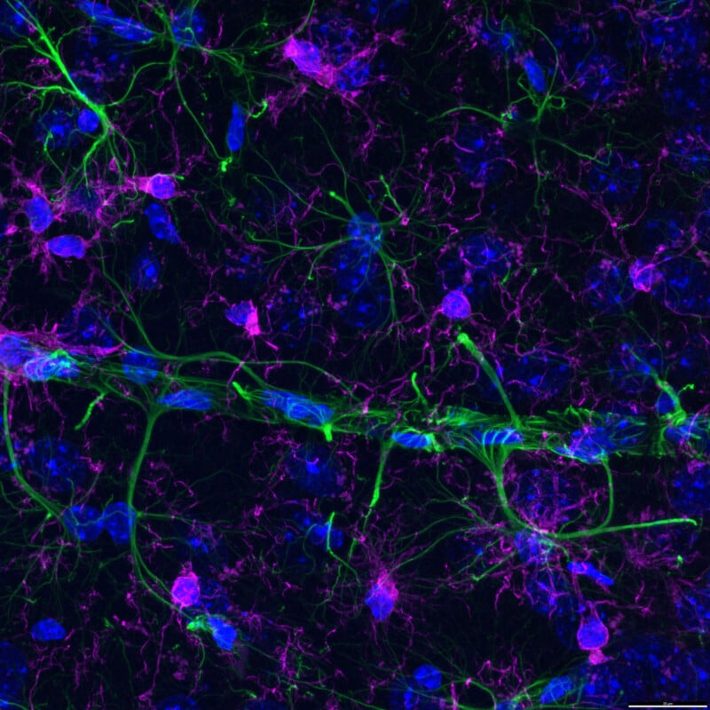
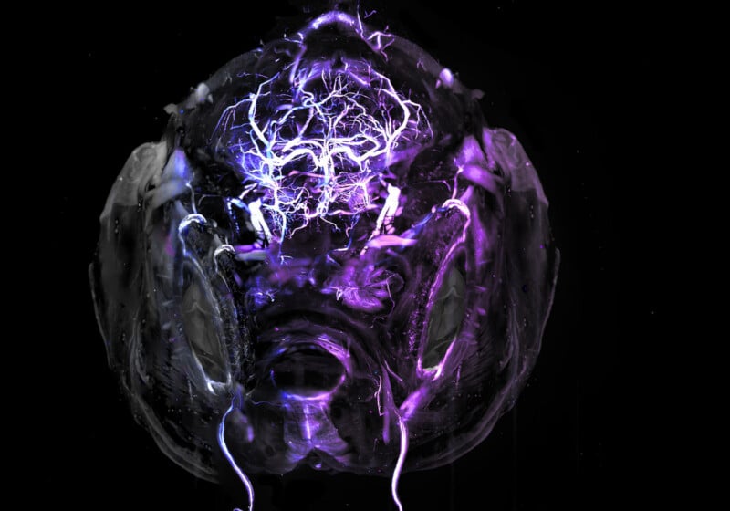
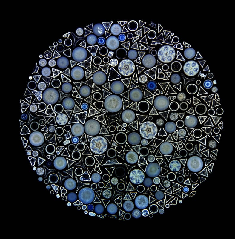
Further, 12 scientific photographers also received honorable mentions for their entries, and each is featured below. 


Fluorescent microscopy image showing colorful brain tissue; orange and purple cell nuclei on the sides, with green neuron projections extending toward the center, and densely packed white cells forming a central vertical band.
© Alexandre Dumoulin
Fluorescent microscopy image of brain cells showing neural structures in green, cell nuclei in blue, and glial cells or processes in magenta, set against a dark background.
© Bettina Rakoczi
A glowing, abstract, purple and white digital illustration resembling a robotic or alien face with electric patterns and energy streaks radiating from the center, set against a black background.
© Hannah Somers
A circular arrangement of variously sized blue and silver circles and triangles, resembling abstract cells or microscopic organisms, set against a black background.
© Jan Rosenboom
Close-up of a jumping spider’s face with multiple eyes, covered in fine hair and yellow pollen grains, against a dark background.
© Javier Ruperez
Colorful microscopic image of a cell with a bright yellow circular outline, a pink nucleus in the center, and blue filaments radiating outward on a dark background.
© Joe McKellar
Black neurons with long, branching dendrites are visible against a pink background, highlighting the complex structure of neural cells. The image resembles a brain tissue section seen under a microscope.
© Marko Pende
Multicolored, fluorescent image of a tiny aquatic organism, possibly a larval crustacean, showing translucent body structures, legs, antennae, and large eyes on a black background.
© Tong Zhang
Extreme close-up of a spider’s eyes, showing two large, glossy, greenish orbs surrounded by fine, brown and tan hair-like structures. The surface of the eyes appears smooth and reflective.
© Walter Ferrari
Colorful, translucent cell-like structures overlap and cluster against a dark background, creating a vibrant, abstract pattern with shades of red, pink, yellow, green, and blue.
© Yue Rong Tan
Close-up image of an insect leg, likely a mosquito, resting on a woven synthetic mesh. The fine hairs and intricate hooks on the leg and detailed threads of the mesh are clearly visible.
© Hange Du
A close-up of a small insect with long, thin legs, antennae, and a feathery tail, set against a black background. The insect’s body appears yellowish with brown segments.
© Hanyang Xue

“We are deeply inspired by the creativity and technical mastery reflected in this year’s entries,” says Wes Pringle, Evident CEO. “Each year, this contest celebrates what’s possible when art and science come together to illuminate the unseen.”

Readers can learn more about how each of the winning photos above was captured, including the specialized equipment each photographer used, on Evident’s website.


Image credits: Evident Scientific. Photographers are credited in the individual captions.

Discussion