Human Hair Knotted With Horsehair One of the Winners of Evident Image of the Year

Left side: A butterfly shape made of colorful, irregular fragments on a black background. Right side: A close-up of a metallic knot against a beige backdrop, featuring two intertwined cylindrical rods.
Photo credit: Jianguo Mao, left, Gerd Günther, right.

The winners of the Evident Image of the Year awards have been announced, a competition that recognizes the world’s best scientific microscopic imaging.

A beautiful image of a Cosmic Orange aster flower taken on a confocal microscope by Igor Siwanowicz has been selected as the global winner.
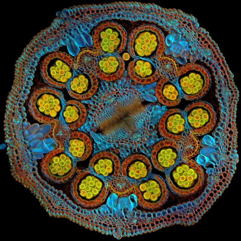
A detailed and colorful microscopic image of a plant stem cross-section. The vibrant pattern features a central area with radiating circular structures in yellow, surrounded by blue and red layers, showcasing its intricate cellular arrangement.
Global Winner. Pollen grains mature inside anthers on a Cosmic Orange Mexican aster flower. | Igor Siwanowicz (USA)

Siwanowicz, a research scientist at the HHMI Janelia Research Campus, picked the flower bud on a post-lunch walk around the campus pond. He says the image “shows that the beauty of a common flower that most of us take for granted can extend beyond what we can see with the naked eye.”

Siwanowicz will receive an Olympus SZX7 stereo microscope with a DP23 digital camera or a set of X Line™ objectives.

Close-up of two intertwined metallic rods forming a secure knot. The rods have a textured surface and a golden-brown appearance, set against a soft beige background.
Materials Science Winner. The image depicts a human hair knotted with horsehair, created with incident brightfield illumination to generate a shimmering metallic effect. The photographer took several attempts to tie the knot for the image. | Gerd Günther (Germany)
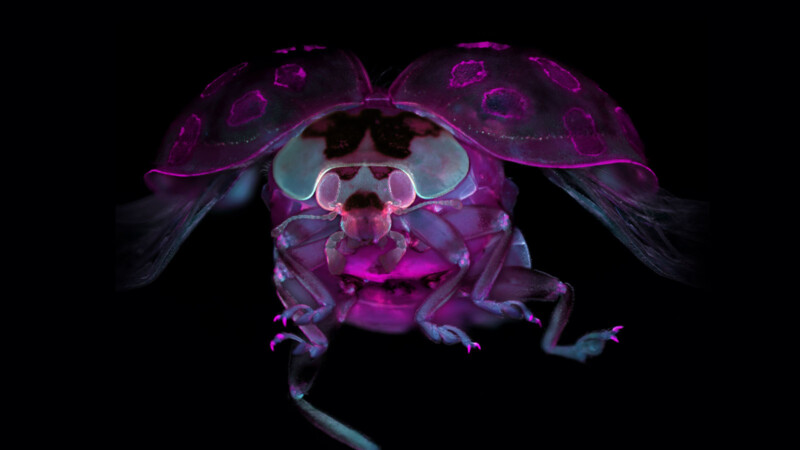
Close-up view of a bright pink and purple ladybug under UV light against a black background. The vivid colors highlight its detailed features, including legs and antennae, creating a vibrant and surreal appearance.
Regional winner for the Americas. Tissue-cleared ladybug. | Marko Pende (USA).
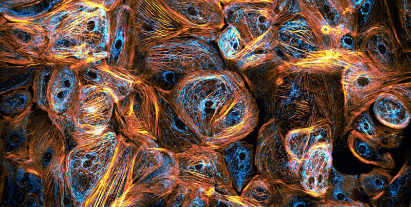
A close-up of colorful cells under a microscope, featuring a network of blue rounded structures and bright orange filamentous patterns against a dark background.
Regional winner for Europe, the Middle East, and Africa. Heart cells derived from induced pluripotent stem cells. | Till Stephan (Germany).
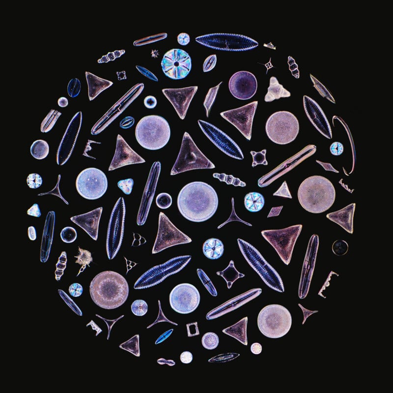
A collection of variously shaped and colorful diatoms is arranged in a circular pattern on a black background, showcasing intricate geometric forms and vibrant iridescent hues.
Regional winner for Asia-Pacific. Diatom arrangement. | Daniel Han (Australia).
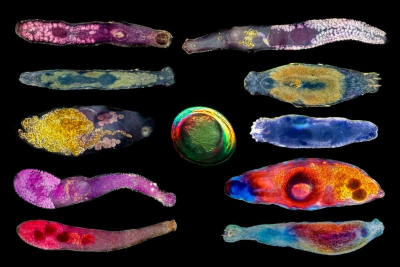
A collage of eleven flatworm specimens displayed against a black background. Each flatworm has distinct colors and patterns, ranging from purples and reds to blues and yellows, with one round, iridescent object in the center.
Honorable Mention. Collage of different fish parasites observed in a dark field in Argentine Patagonia. | Antonio Segura & Priscilla Vieto (Argentina)
An arrangement of colorful pills on a black background forming the shape of a butterfly.
Honorable Mention. Butterfly image consists of more than 200 scale patterns taken from different butterfly wings. | Jianguo Mao (China)
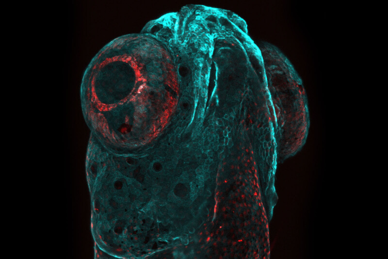
Microscopic image of a zebrafish larva's head, showing large, round eyes. The image is colored with a blue and red overlay, highlighting the detailed cellular structures against a black background.
Honorable Mention. The submarine. The image is a depict of a zebrafish head covered by epithelial cells expressing the palm-mTurquoise fluorescent protein and mCherry nuclear protein. | Yue Rong Tan (Taiwan)
Colorful neon light strips arranged in abstract shapes against a dark background. A glowing orb with a pink and blue aura is visible above, adding a cosmic touch to the scene.
Honorable Mention. Fluorescent images of normal rod-shaped filamentous cells of the common soil bacterium Bacillus subtilis and a spherical cell representative that has lost its cell wall. | Sandra Story (USA)
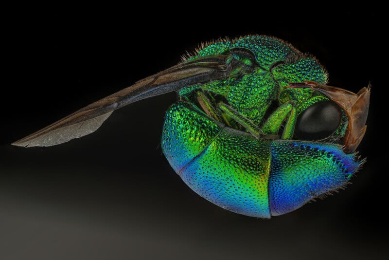
Close-up image of a vividly colored insect with iridescent green and blue scales on its body and a large, black eye set against a dark background. The detailed texture of the insect's body and wings is clearly visible.
Honorable Mention. A cuckoo wasp. Cuckoo wasps are captivating insects, showcasing a dazzling array of iridescent colors. | Raghuram Annadana (India)
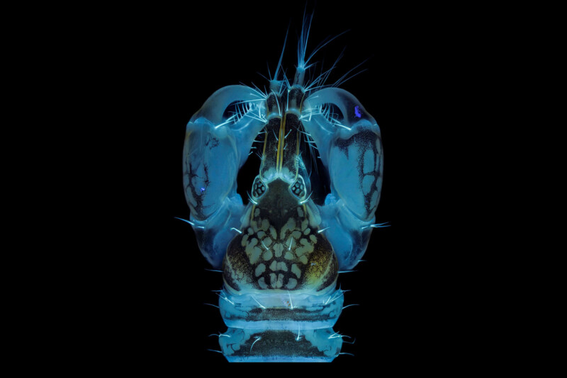
Close-up of a colorful, luminescent jumping spider set against a black background. The spider displays vibrant blue and green hues with intricate patterns and fine hairs, creating an otherworldly and detailed appearance.
Honorable Mention. Tanaidacea crustacean swims in the water column during night and is attracted by light. Chitin emit light when excited using UV light. | Luigi Olivo Bozzano (Italy)
Close-up of a vibrant, colorful sea creature resembling a starfish, with multiple arms covered in spiky, intricate patterns. The colors include bright pink, purple, and orange against a black background.
Honorable Mention. Juvenile sea star skeleton. Sea star (Patiria miniata) juvenile stained with calcein and DAPI. | Laurent Formery (USA)
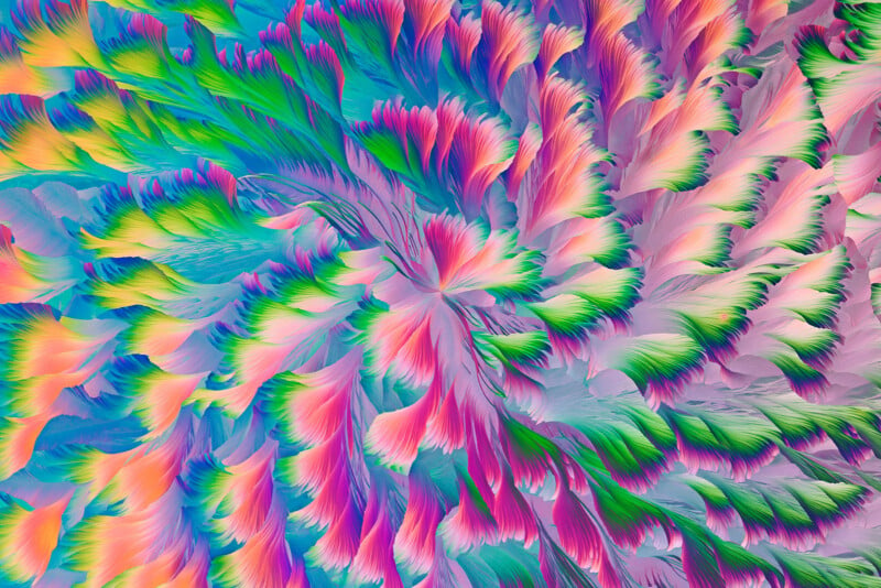
Abstract image featuring a vibrant, swirling pattern of multicolored feather-like shapes. The design includes shades of pink, blue, green, and purple, creating a dynamic and colorful composition.
Honorable Mention. Crystalline pattern, like the big cotton padded jacket pattern handed down during the Chinese Spring Festival. | Ji Yuan (China)
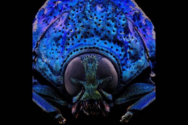
Close-up image of a vibrant blue and purple beetle with intricate black spots and large, detailed eyes on a black background, highlighting the beetle's textured exoskeleton.
Honorable Mention. A warty leaf beetle (Poropleura) that looks like a prehistoric tyrannosaurus. | Hanyang Xue (China)
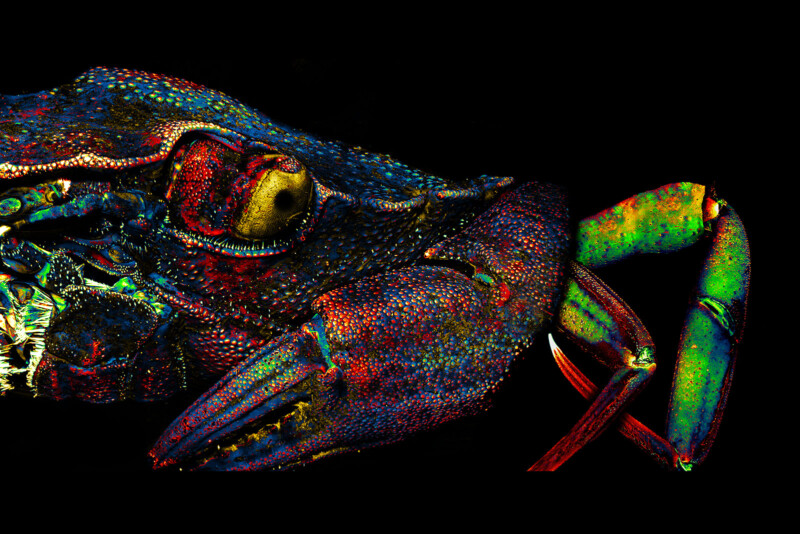
Close-up of a crab in vibrant colors against a black background. The image highlights detailed features, including textured skin with red, blue, and green hues, prominent eye, and sharp pincers, creating a striking and surreal appearance.
Honorable Mention. Green crab, labeled with DAPI and imaged autofluorescence, reflected light. | Hannah Somers (USA)
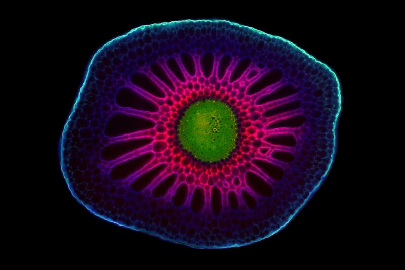
Microscopic image showing a brightly colored cross-section of a plant stem. The outer layer is a glowing blue, surrounding a vivid pink area with polyp-like structures radiating from a central green core, all against a black background.
Honorable Mention. A look into the depths. Cross sections through the apical stem segments of the aquatic species Utricularia macrorhiza. | Hana Sehadová (Czech Republic)
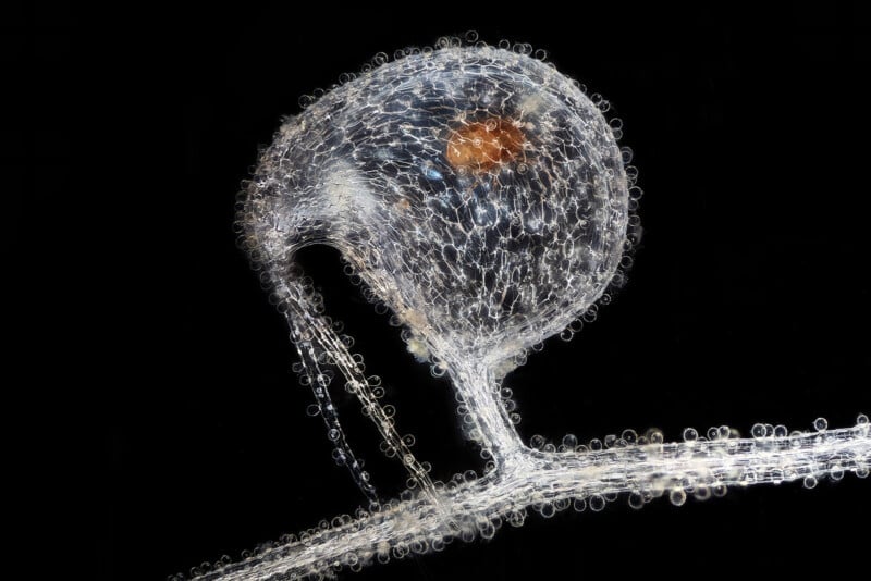
Close-up of a transparent aquatic organism against a black background, featuring a bulbous head with an internal brownish structure. The body extends into a long, thin tail-like appendage with a series of small, spherical bubbles.
Honorable Mention. Trap of subaquatic carnivorous bladderwort Utricularia humboldtii with trapped water mite Hydrachnidia. | AnFrantisek Bednar (Slovakia)

Evident came about after the Olympus Corporation spun off its Scientific Solutions Division in 2022 to form a new company. Evident’s Image of the Year Award began as the Image of the Year European Life Science Light Microscopy Award with the aim to celebrate both the artistic and scientific value of microscopy images.

For more information, visit EvidentScientific.com. The competition can be viewed here.

Discussion