Olympus Unveils the Winners of the Best Life Science Photos of 2020
![]()
Olympus has announced the winners of its Global Image of the Year Life Science Light Microscopy Award, which is an annual competition that recognizes the best in life science photos taken with a microscope.
This competition is hosted by Olympus’s Life Sciences division, which is not to be confused with the newly-formed OM Digital under Japan Industrial Partners. The competition started in 2017 as the Image of the Year European Life Science Light Microscopy Award with the aim to celebrate both the artistic and scientific value of microscopy images. Today, the organizers say that the competition stays true to this mission by encouraging people across the world to look at scientific images in a new way, appreciate their beauty, and share images with others.
To be considered for this year’s competition, images had to be submitted between September 15, 2020 and January 31, 2021. A maximum of three images was allowed per participant, and anyone over the age of 18 could participate (provided that they are not an Olympus employee, family member, judge, or family member of a judge nor engaged in the fields of manufacturing or sales of microscopes).
All entries were evaluated on artistic and visual aspects, scientific impact, and microscope proficiency.
From rat embryos to butterfly scales and snakeskin, the Olympus Life Science team was impressed by the diverse collection captured under the microscope this year.
This year’s competition saw nearly 700 submissions from 61 different countries, with the winning image going to Werner Zuschratter from Germany. Zuschratter submitted the incredible photo below depicting a whole rat embryo that he captured with a confocal microscope. For the grand prize, Zuschratter will receive an Olympus SZX7 stereo microscope with a DP27 digital camera.
![]()
In addition to the overall global winner, three regional awards were presented to XinPei Zhang (China) for Asia, Justin Zoll (USA) for the Americas, and Grigorii Timin (Switzerland) for EMEA. Each regional winner will receive an Olympus CX23 upright microscope.
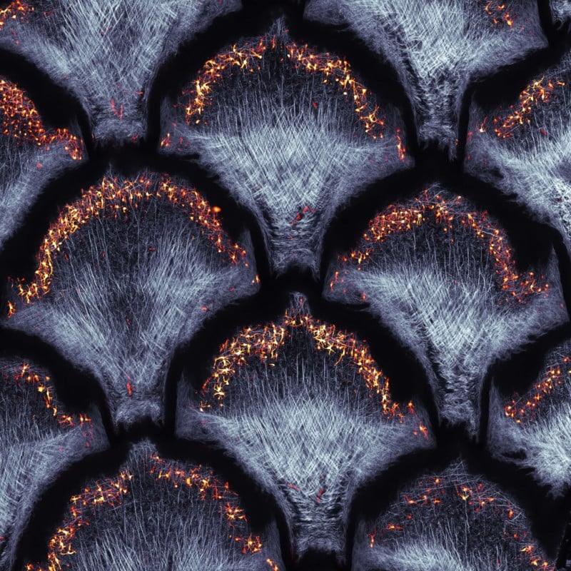
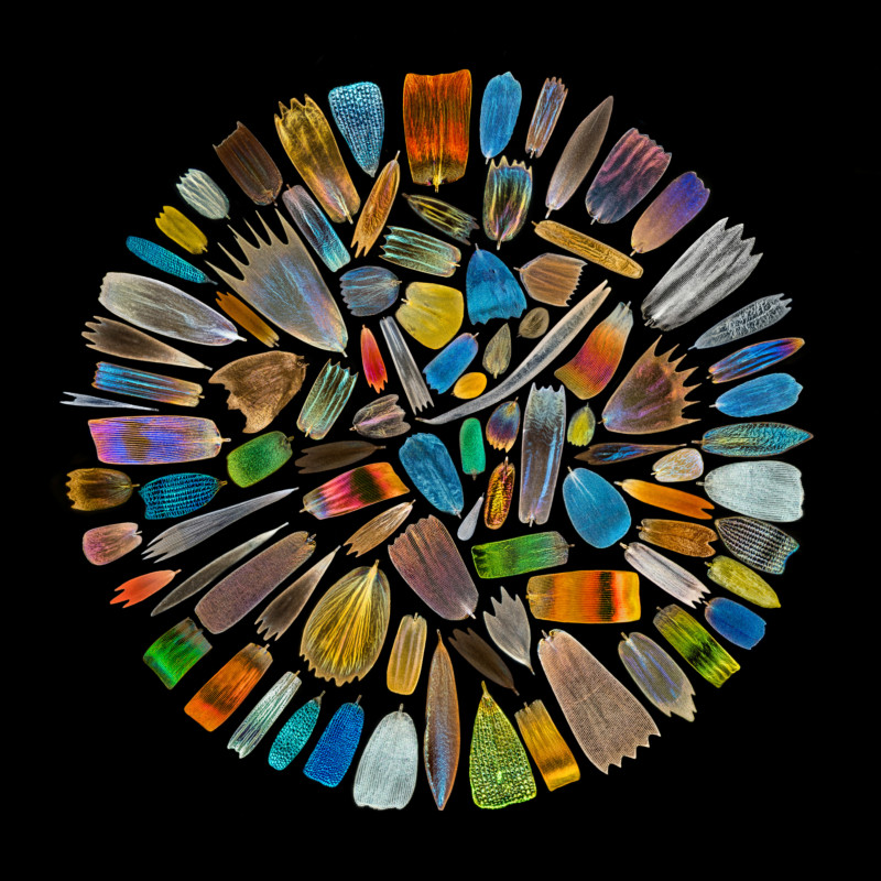

Though they may not have brought home the grand prize or won in any of the three regions, the honorable mentions below are still breathtaking images.
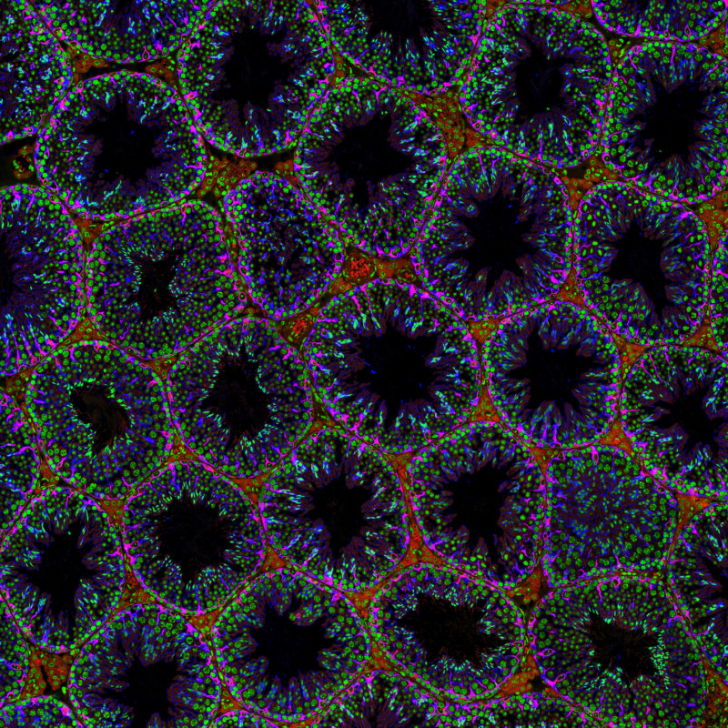
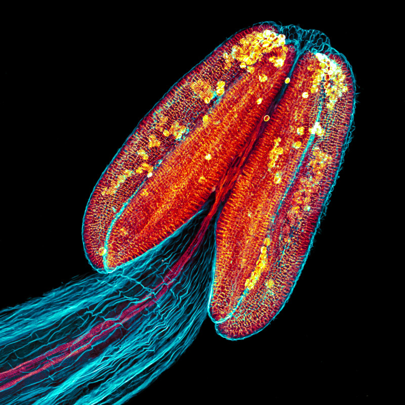
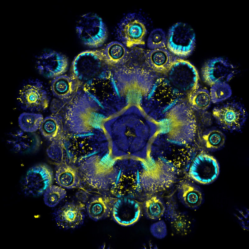
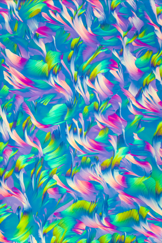
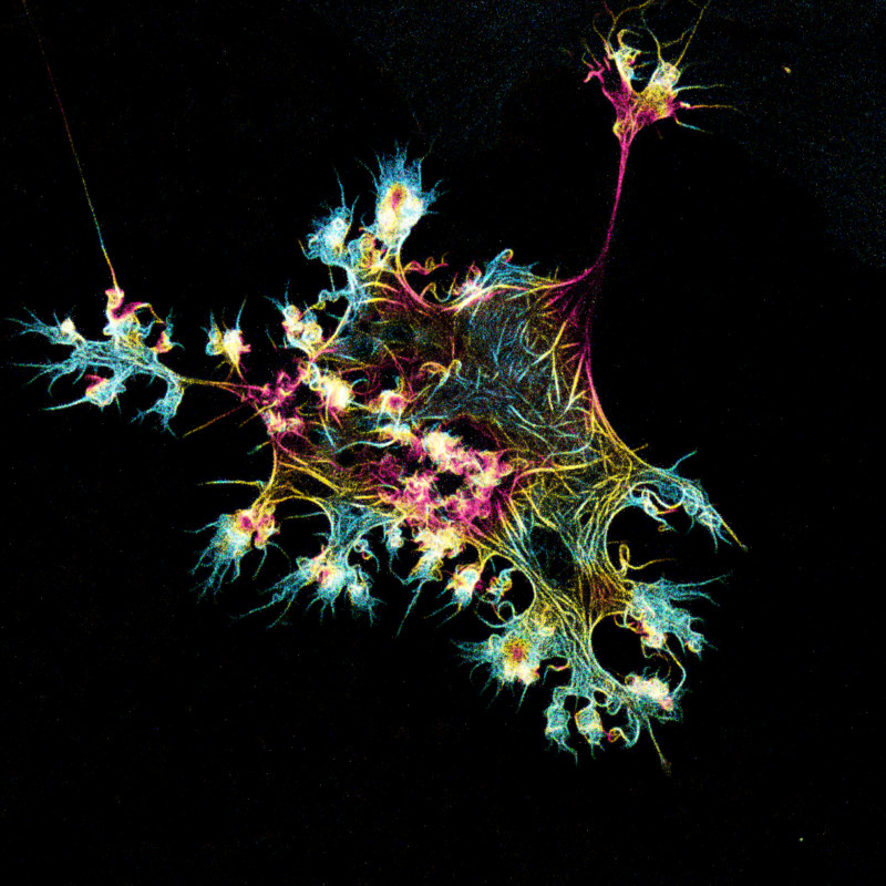
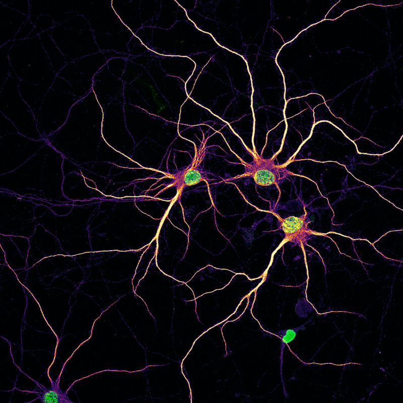
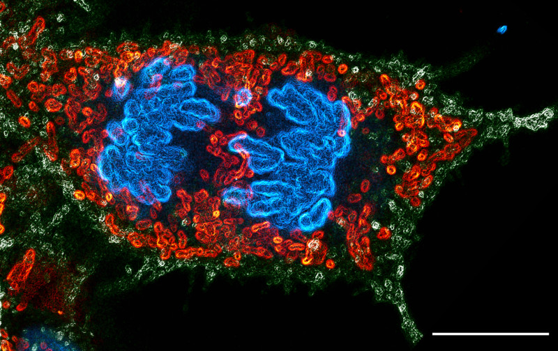
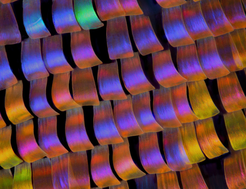
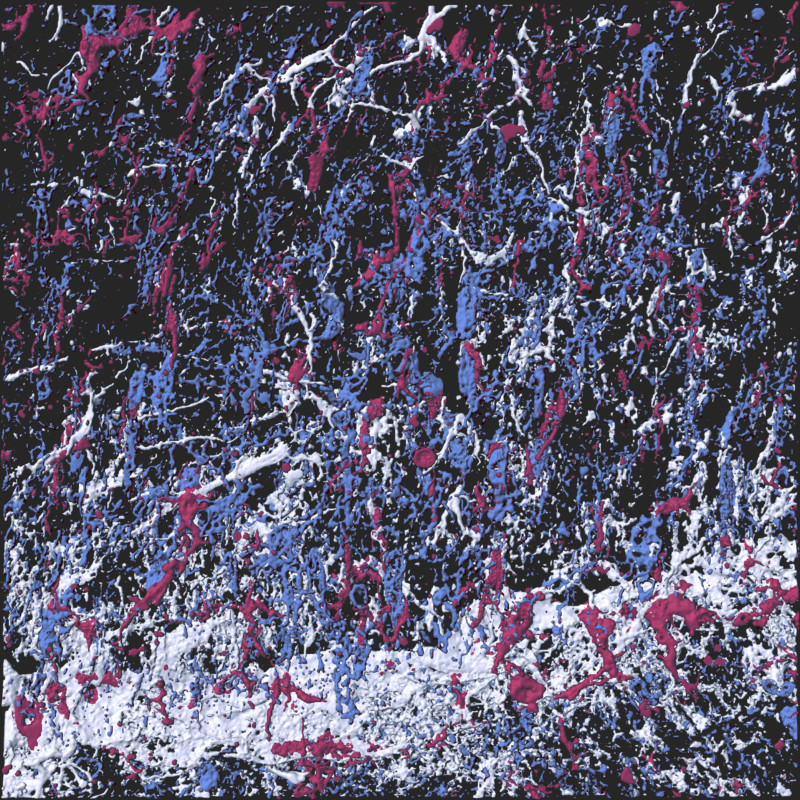
To read about the jurors and see past competition winners, visit the Olympus Lifescience website here.
Image credits: All photos individually credited and provided courtesy of Olympus Life Science.