Photo of a Rodent’s Optic Nerve Wins Nikon’s 2023 Small World Competition
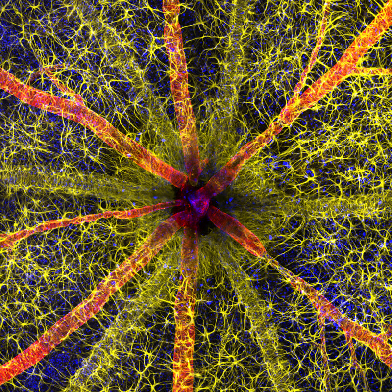
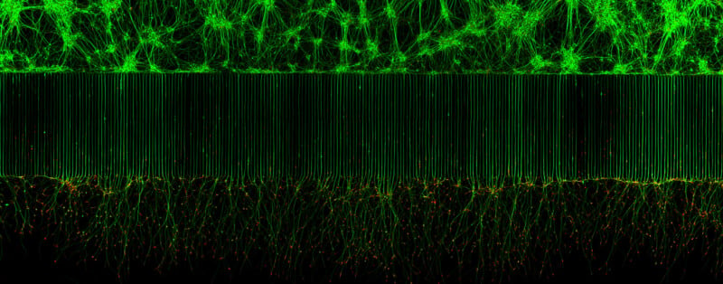
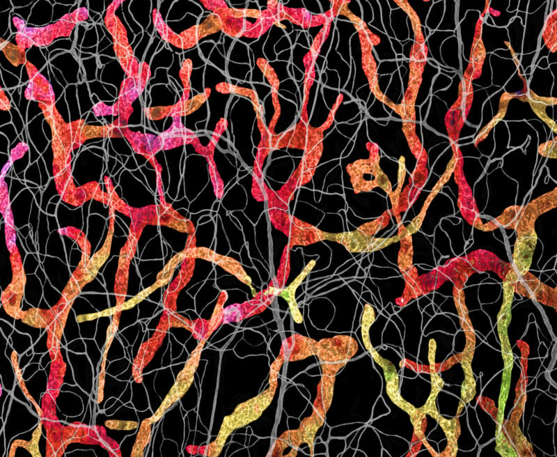
Nikon’s 49th annual Small World Photomicrography Competition announced its winners and the top prize went to Hassanain Qambari for his vivid image of a rodent optic nerve head.
The Nikon Small World Competition first began in 1975 as a means to recognize the efforts of those involved with photography through the light microscope. Since then, Small World has become a leading showcase for photomicrographers that Nikon says includes work from the widest array of scientific disciplines. This year, the competition received nearly 1,900 photo entries from 72 countries.
The photo competition results follow the video results — called Nikon Small World in Motion — that were revealed in September.
Winning images are judged on originality, informational content, technical proficiency, and visual impact.
The winning photo above, which was captured by Qambari and assisted by Jayden Dickson of the Lions Eye Institute, shows extreme detail, including the astrocytes (yellow), contractile proteins (red), and retinal vasculature (green) from inside the optic nerve head. Nikon explains that the colorful image provides an important contribution to the study and reversal of diabetic retinopathy, which affects one in five persons with diabetes worldwide.
“Diabetic retinopathy occurs when high blood sugar damages the blood vessels in the tissue at the back of the eye, known as the retina. The damaged blood vessels can swell and leak, which can cause blurry vision or total loss of eyesight. Since 2021, Qambari has devoted his time and research to the early detection and reversal of the disease,” Nikon explains.
“Current diagnostic criteria and treatment regimens for diabetic retinopathy are limited to the late-stage appearance of the disease, with irreversible damage to retinal microvasculature and function,” Qambari says. “The visual system is a complex and highly specialized organ, with even relatively minor perturbations to the retinal circulation able to cause devastating vision loss. I entered the competition as a way to showcase the complexity of retinal microcirculation.”
Nikon says that Qambari’s efforts to capture the winning image weren’t without challenges, not the least of which was locating the fine vessels that are nearly 110 microns in diameter.
“Over the past 20 years, our research group has refined the technique of isolated ocular perfusion labeling for fine vessels in the eye,” Qambari explains. “While the ophthalmic artery in the rodent model presented a technically demanding challenge, we were able to overcome it with persistence and patience.”
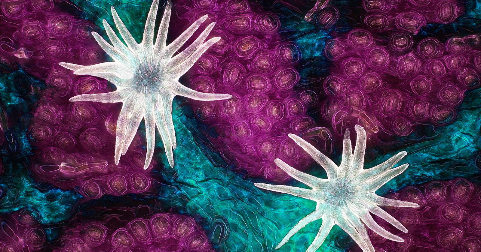
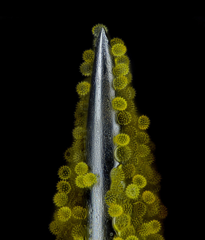
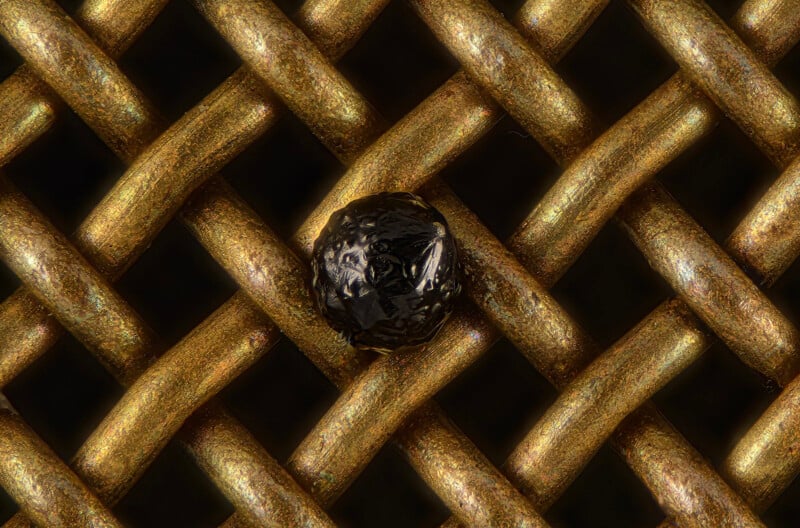
Second place was awarded to Ole Bielfeldt for his image of a matchstick igniting by the friction surface of the box. The image was taken within one eight-thousands of a second and utilized imaging stacking.

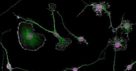
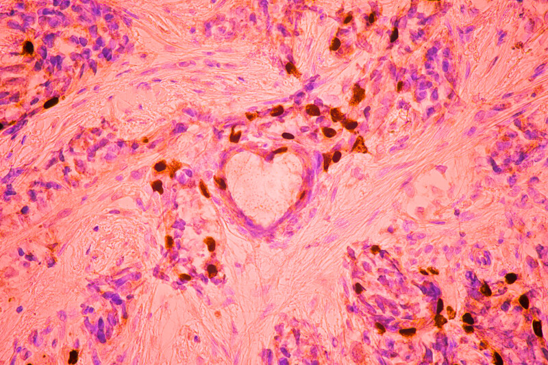
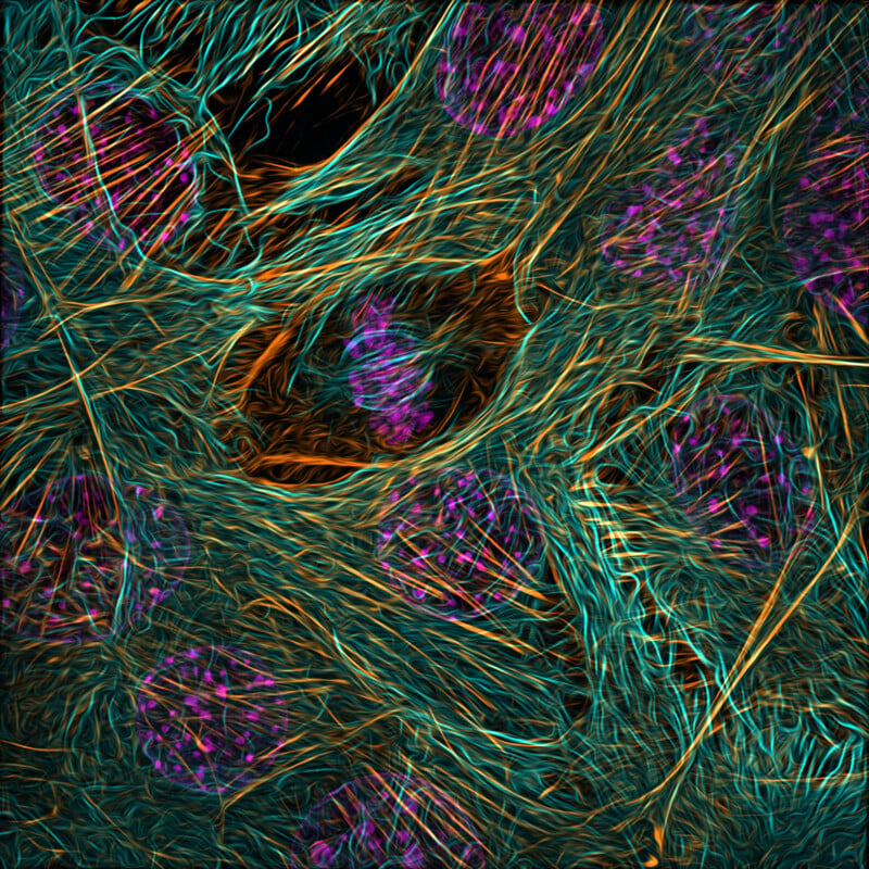
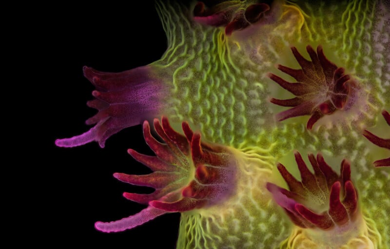
Third place was awarded to Malgorzata Lisowska for her image of breast cancer cells (below).
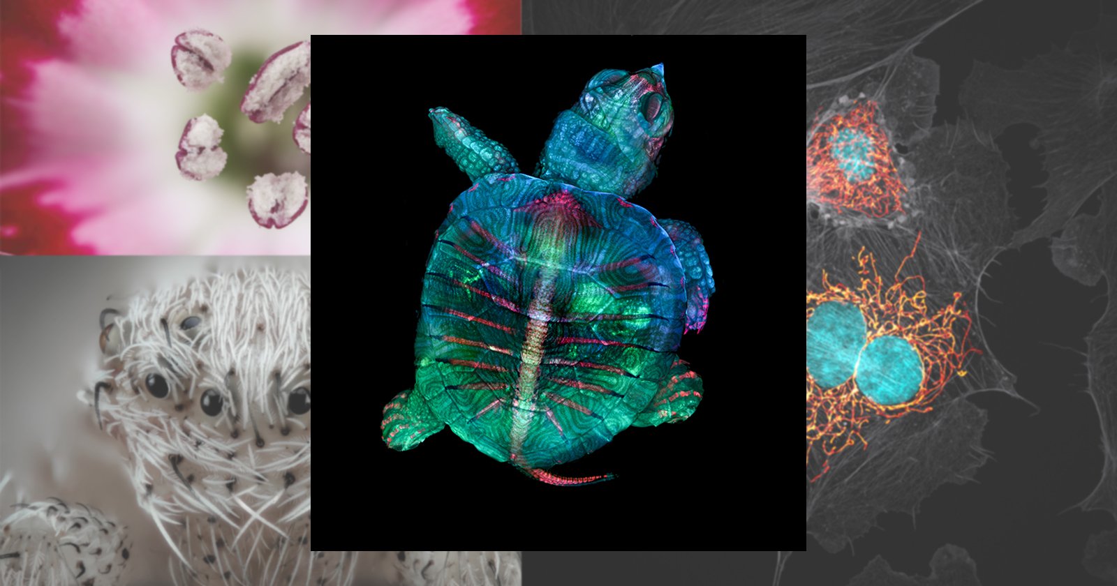
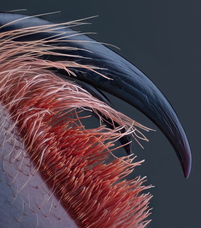
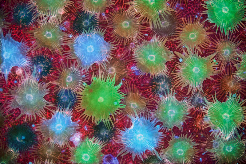
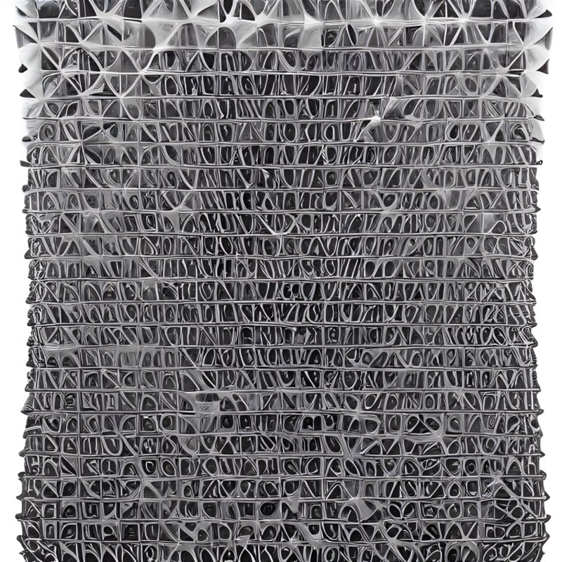
Below are the remaning 17 top awarded finalists, from fourth place through 20th place in order.


In total, Nikon Small World recognized 83 photos out of the thousands it received, all of which can be seen on the competition’s website.
Image credits: All photos are individually credited and provided courtesy of Nikon Small World.