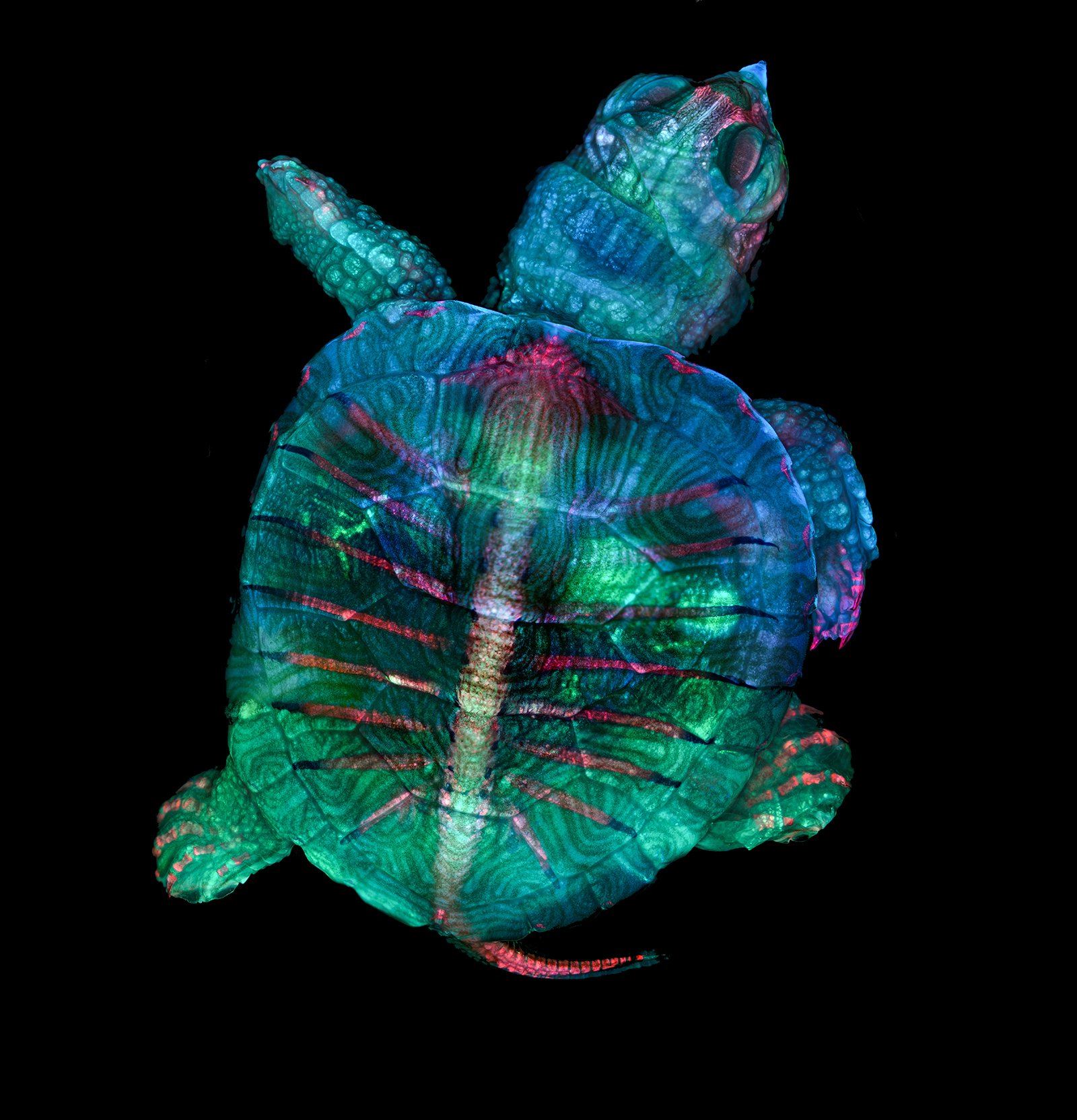
Beautiful Image of Turtle Embryo Wins Nikon Small World Photo Contest
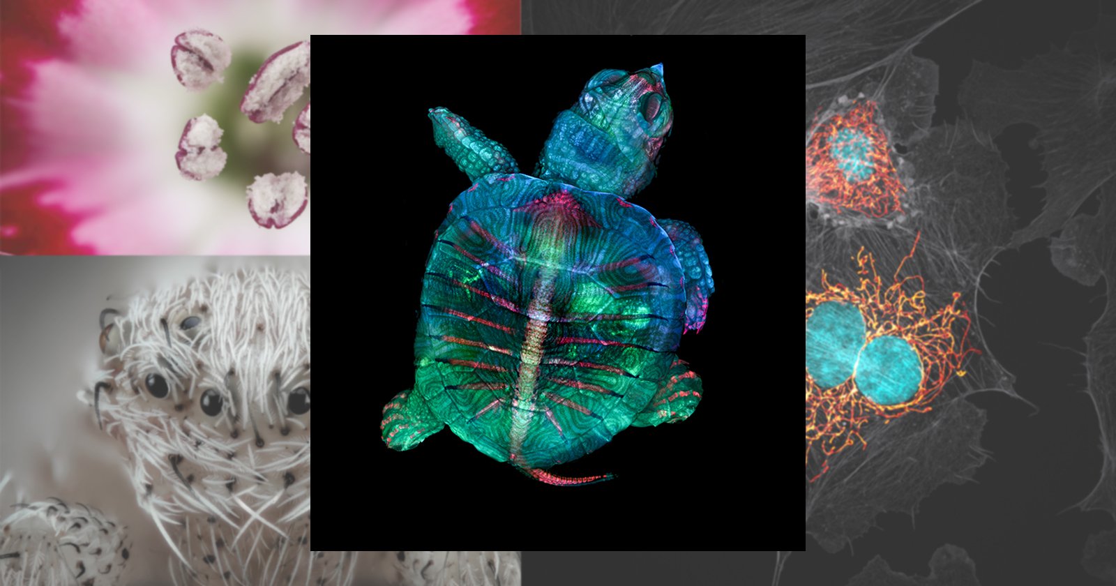
Nikon has revealed the winners of the 45th annual Nikon Small World photomicrography competition. The photo contest features images that “showcase a spectacular blend of science and artistry under the microscope,” and as usual, this year’s winning images are just plain spectacular.
“Microscopy lets us zoom in on the smallest organisms and building blocks that comprise our world – giving us a profound appreciation for the small things in life that far too often go unnoticed,” Kugler told Nikon, “It allows me to do science with a purpose.”
“We are inspired by the beautiful images we see through the microscope,” added Zgoda, “It’s humbling and deeply fulfilling to be able to share that science with other people.”
Of course, Zgoda and Kugler’s photograph isn’t the only incredible image recognized by the contest this year. A “Top 20” were selected, and you can see all of them—including a brief caption that explains what you’re seeing and how it was captured—below.
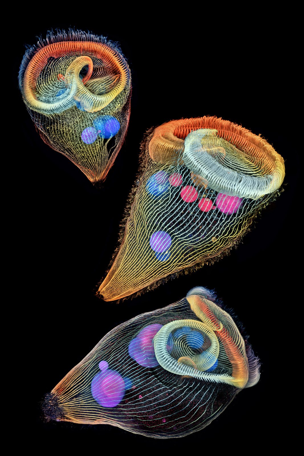
2nd Place – Dr. Igor Siwanowicz
Howard Hughes Medical Institute (HHMI)
Janelia Research Campus
Ashburn, Virginia, USA
Depth-color coded projections of three stentors (single-cell freshwater protozoans)
Confocal
40x (Objective Lens Magnification)
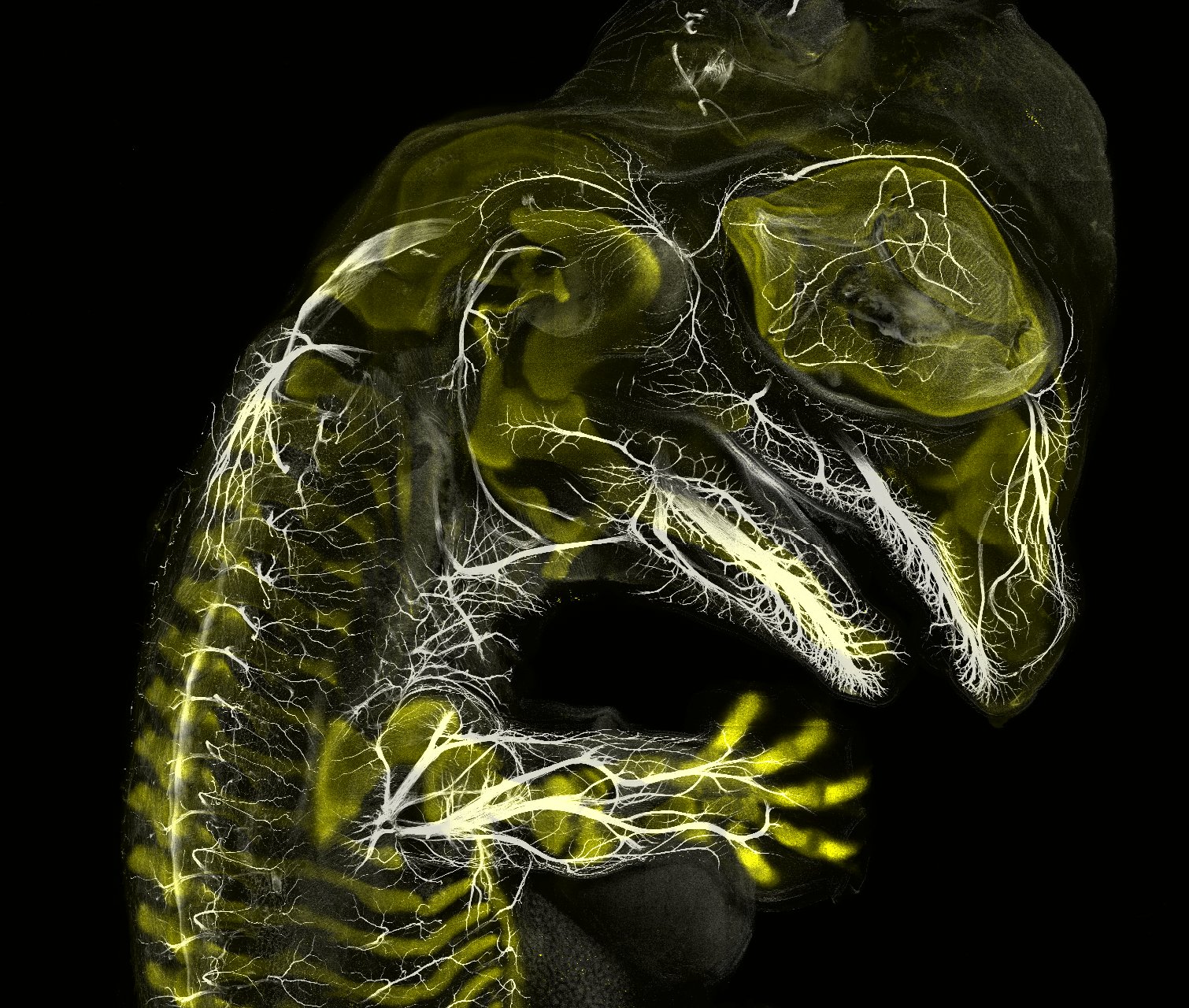
3rd Place – Daniel Smith Paredes & Dr. Bhart-Anjan S. Bhullar
Yale University
Department of Geology and Geophysics
New Haven, Connecticut, USA
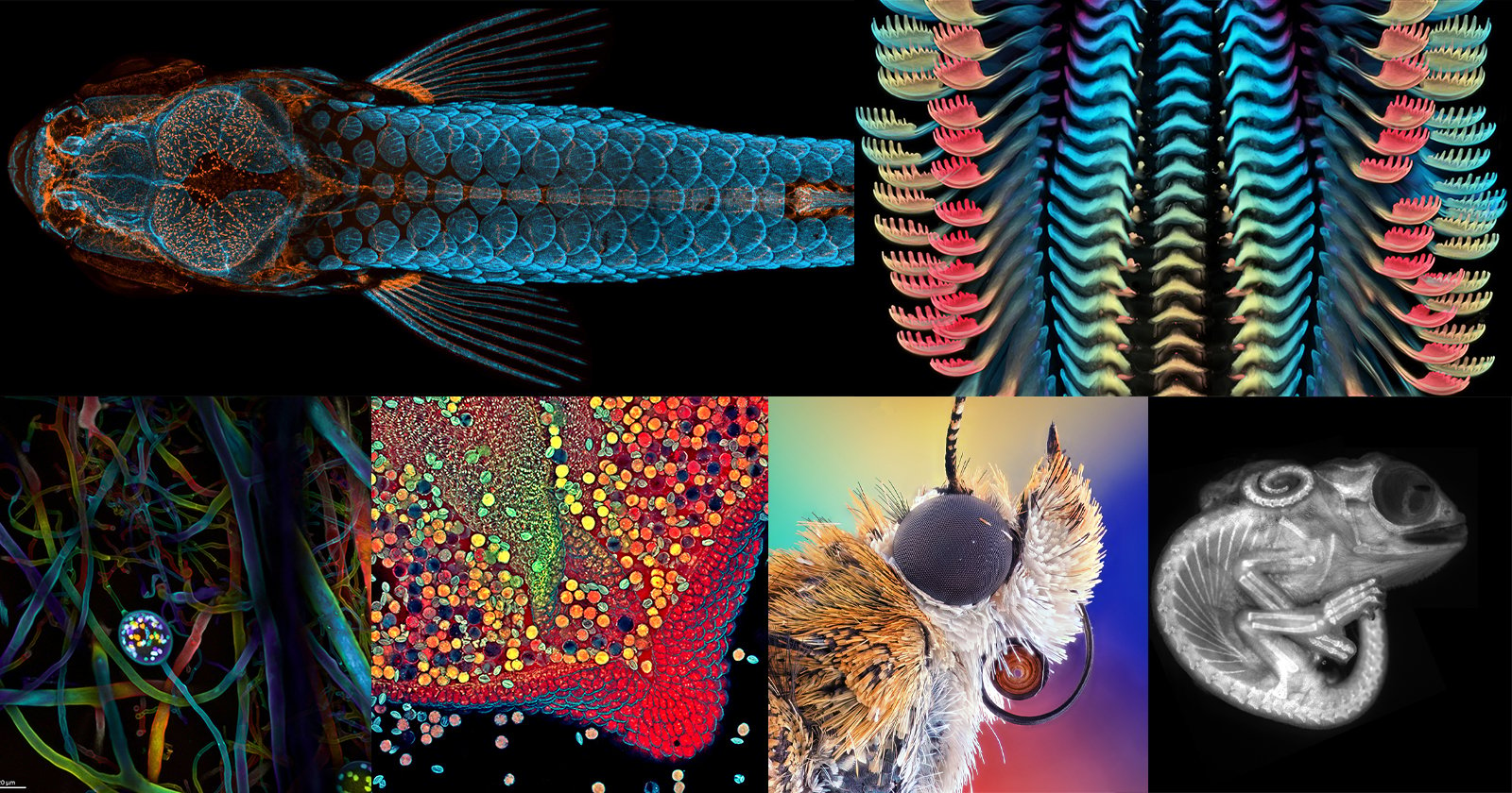
Alligator embryo developing nerves and skeleton
Immunofluorescence
10x (Objective Lens Magnification)
4th Place – Jan Rosenboom

Universität Rostock
Rostock, Mecklenburg Vorpommern, Germany
Male mosquito
Focus Stacking
6.3x (Objective Lens Magnification)
5th Place – Caleb Foster
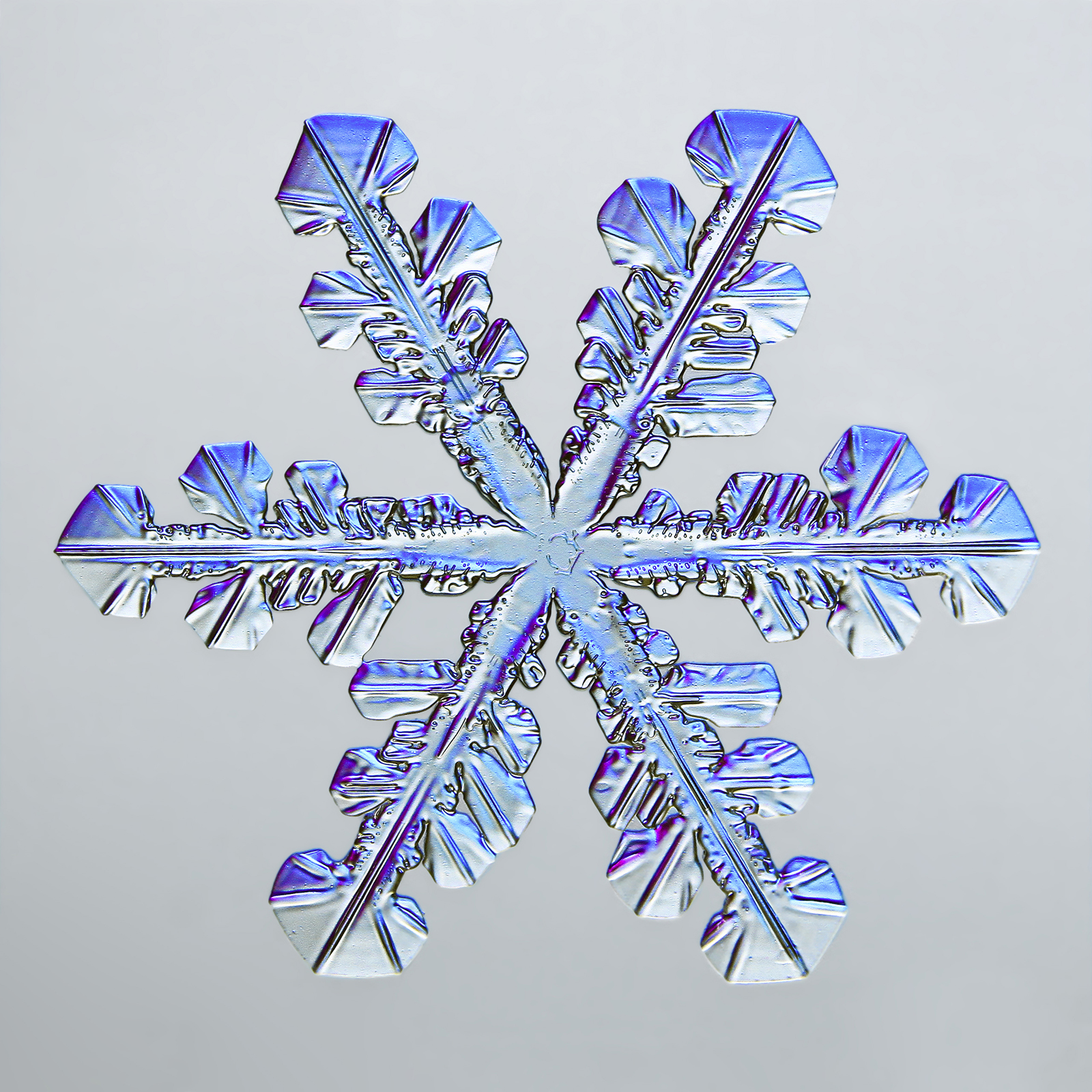
Caleb Foster Photography
Jericho, Vermont, USA
Snowflake
Transmitted Light
4x (Objective Lens Magnification)
6th Place – Javier Rupérez
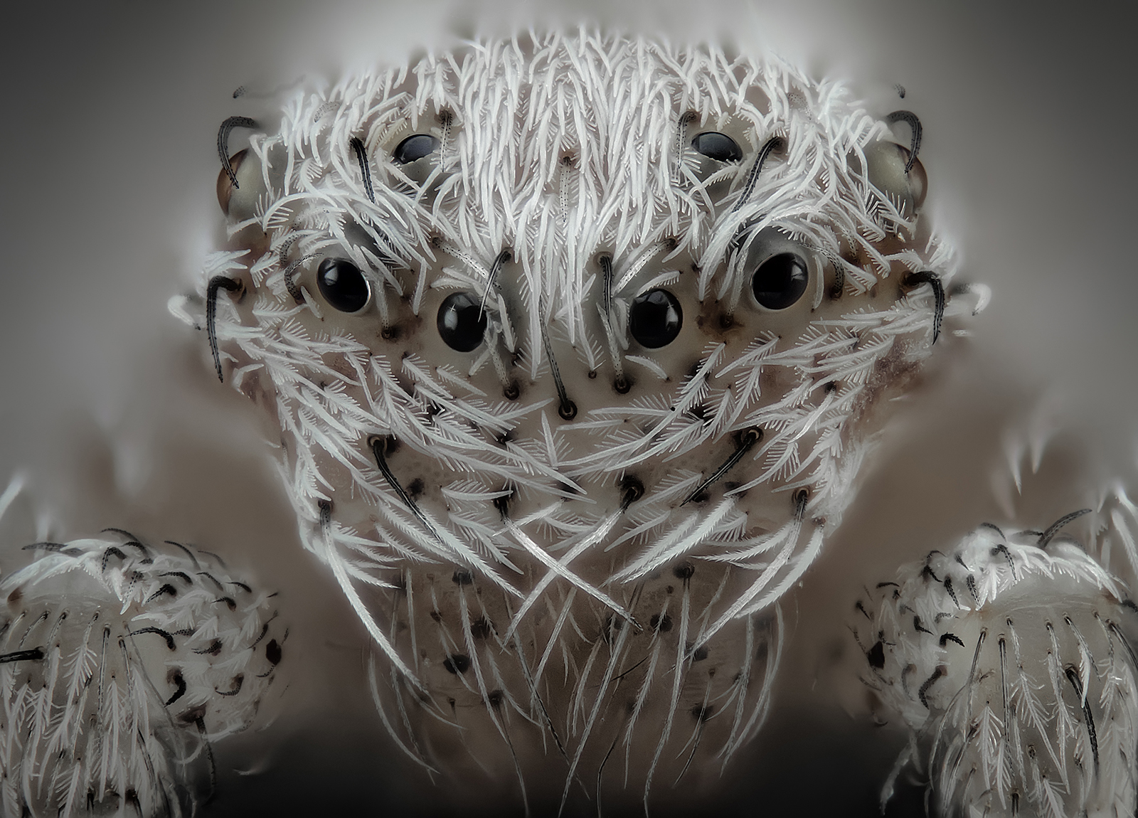
Almáchar, Málaga, Spain
Small white hair spider
Reflected Light, Image Stacking
20x (Objective Lens Magnification)
7th Place – Dr. Guillermo López
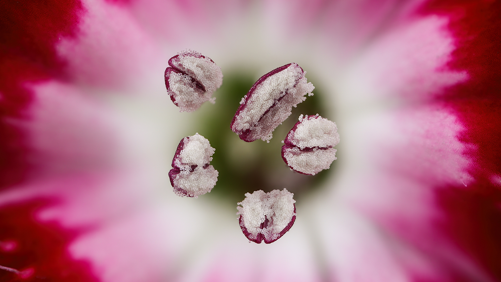
Alicante, Spain
Chinese red carnation stamen
Focus Stacking
3x (Objective Lens Magnification)
8th Place – Garzon Christian

Quintin, Cotes-d’Armor, France
Frozen water droplet
Incident Light
8x (Objective Lens Magnification)
9th Place – Andrei Savitsky
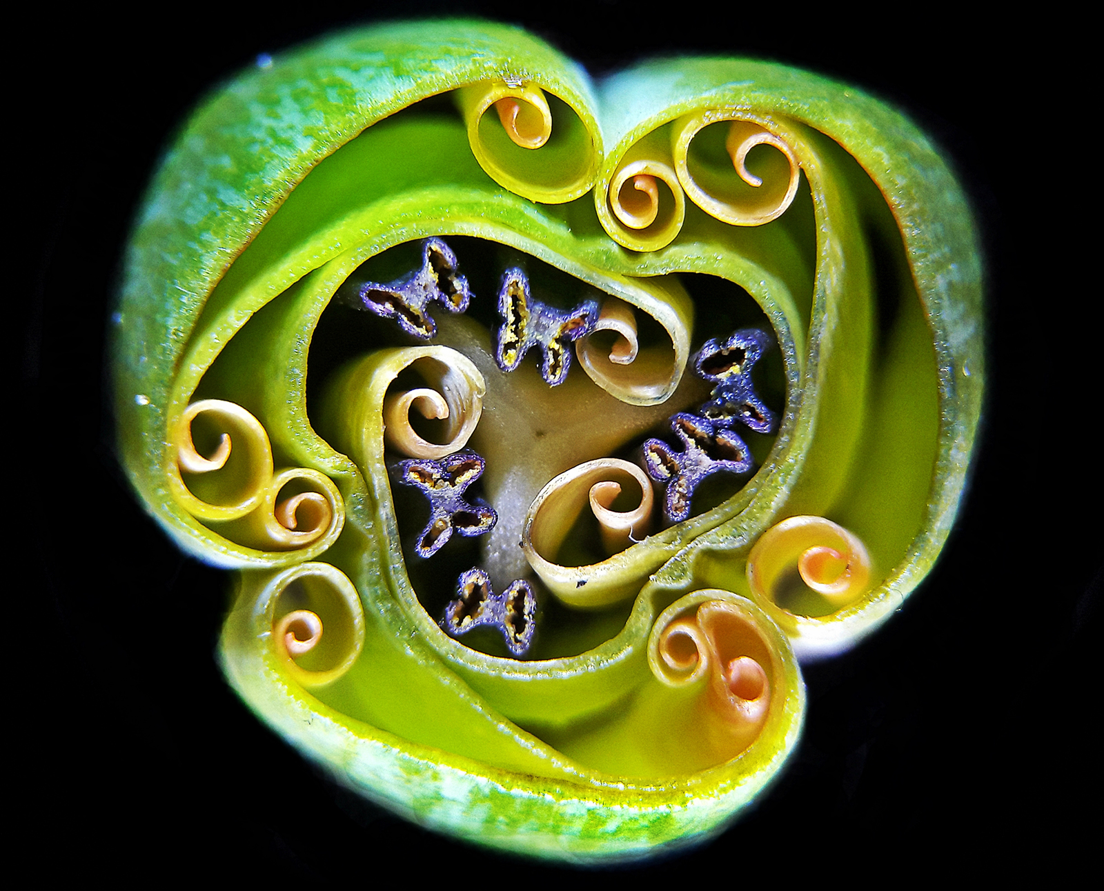
Cherkassy, Ukraine
Tulip bud cross section
Reflected Light
1x (Objective Lens Magnification)
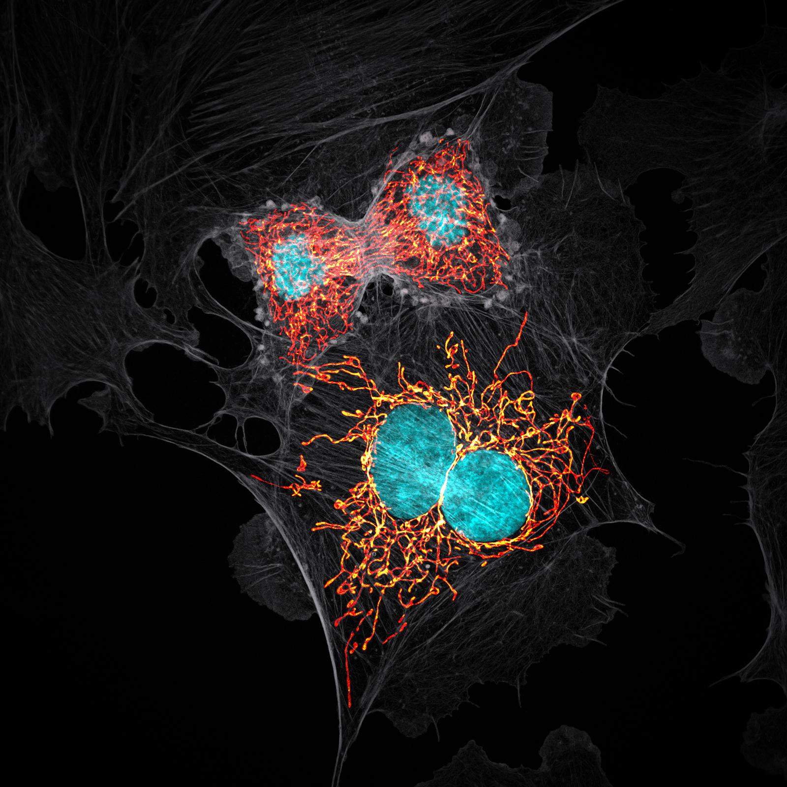
10th Place – Jason M. Kirk
Baylor College of Medicine
Optical Imaging & Vital Microscopy Core
Houston, Texas, USA
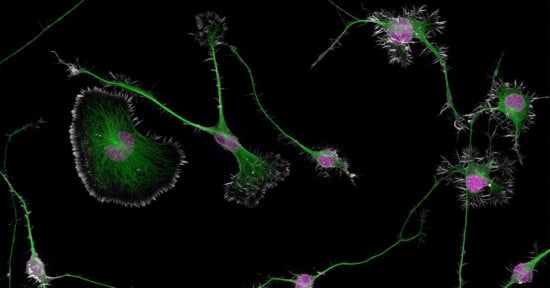
BPAE cells in telophase stage of mitosis
Confocal with Enhanced Resolution
63x (Objective Lens Magnification)
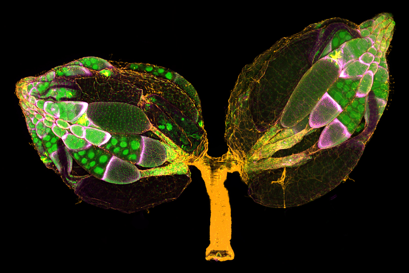
11th Place – Dr. Yujun Chen & Dr. Jocelyn McDonald
Kansas State University
Department of Biology
Manhattan, Kansas, USA
A pair of ovaries from an adult Drosophila female stained for F-actin (yellow) and nuclei (green); follicle cells are marked by GFP (magenta)
Confocal
10x (Objective Lens Magnification)
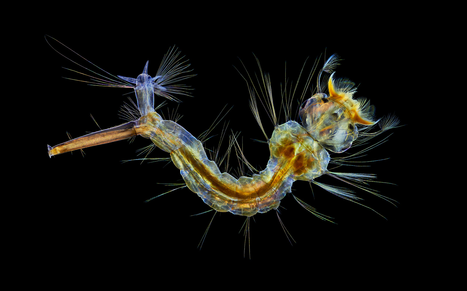
12th Place – Anne Algar
Hounslow, Middlesex, United Kingdom
Mosquito larva
Darkfield, Polarizing Light, Image Stacking
4x (Objective Lens Magnification)
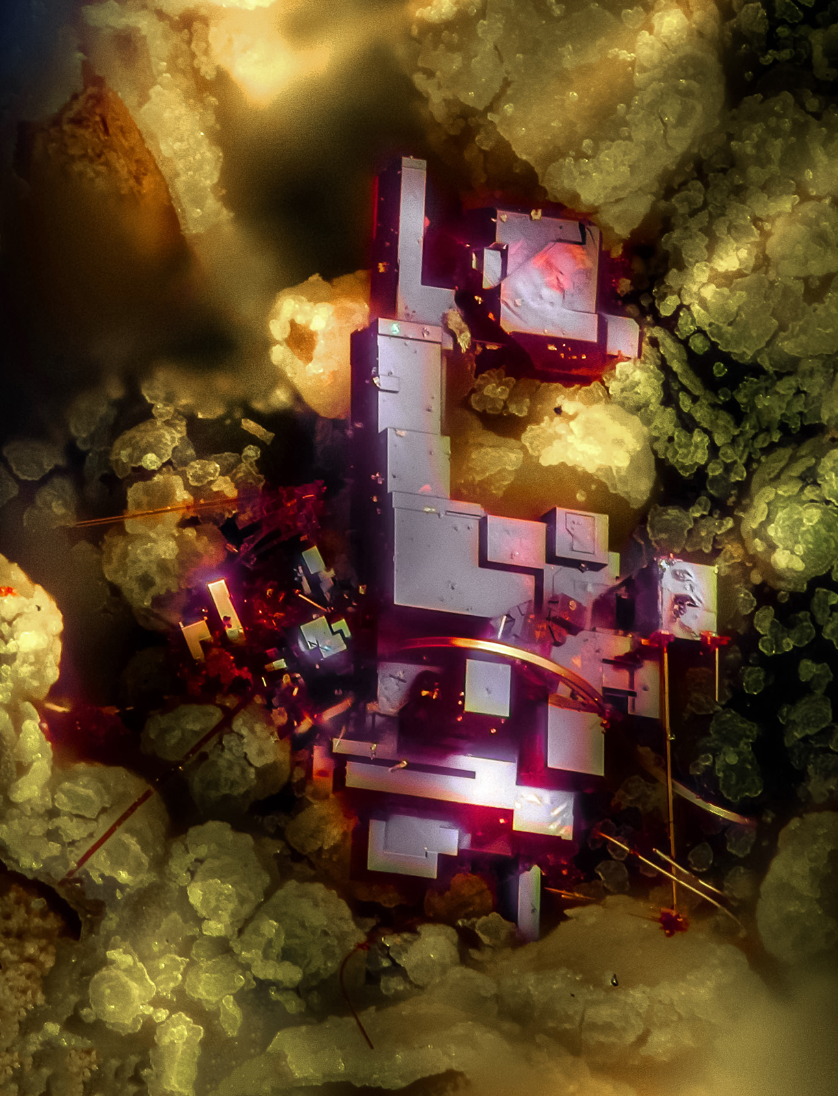
13th Place – Dr. Emilio Carabajal Márquez
Madrid, Spain
Cuprite (mineral composed of copper oxide)
Focus Stacking
20x (Objective Lens Magnification)
14th Place – Antoine Franck
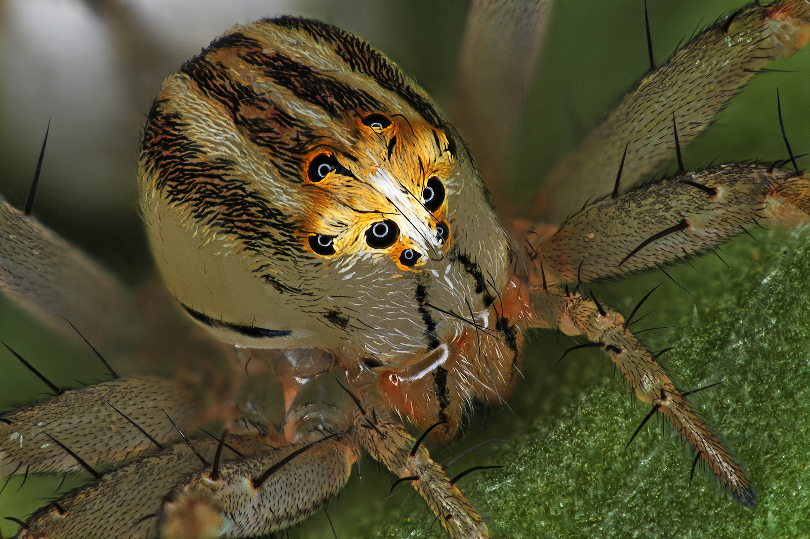
CIRAD – Agricultural Research for Development
Saint Pierre, Réunion
Female Oxyopes dumonti (lynx) spider
Focus Stacking
1x (Objective Lens Magnification)
15th Place – Marek Miś
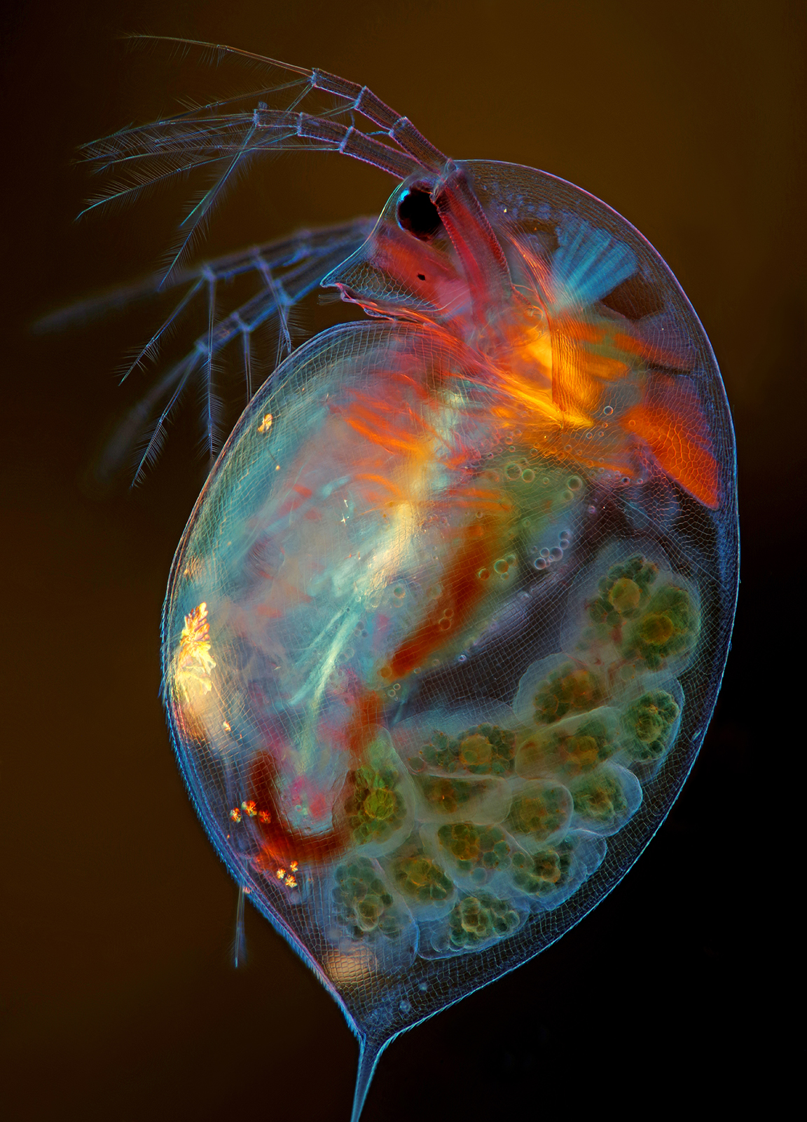
Marek Miś Photography
Suwalki, Podlaskie, Poland
Pregnant Daphnia magna (small planktonic crustacean)
Modified Darkfield, Polarized Light, Image Stacking
4x (Objective Lens Magnification)
16th Place – Dr. Razvan Cornel Constantin
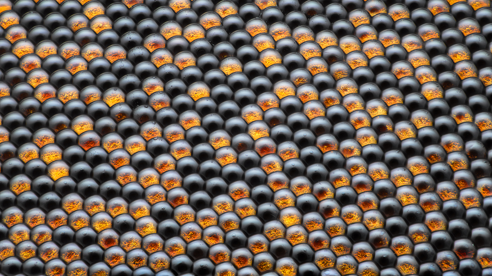
Bucharest, Romania
Housefly compound eye pattern
Focus Stacking, Reflected Light
50x (Objective Lens Magnification)
17th Place – Karl Deckart
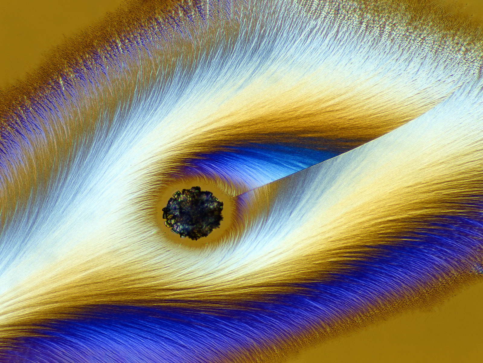
Eckental, Bavaria, Germany
Vitamin C
Brightfield, Polarized Light
4x (Objective Lens Magnification)
18th Place – E. Billie Hughes
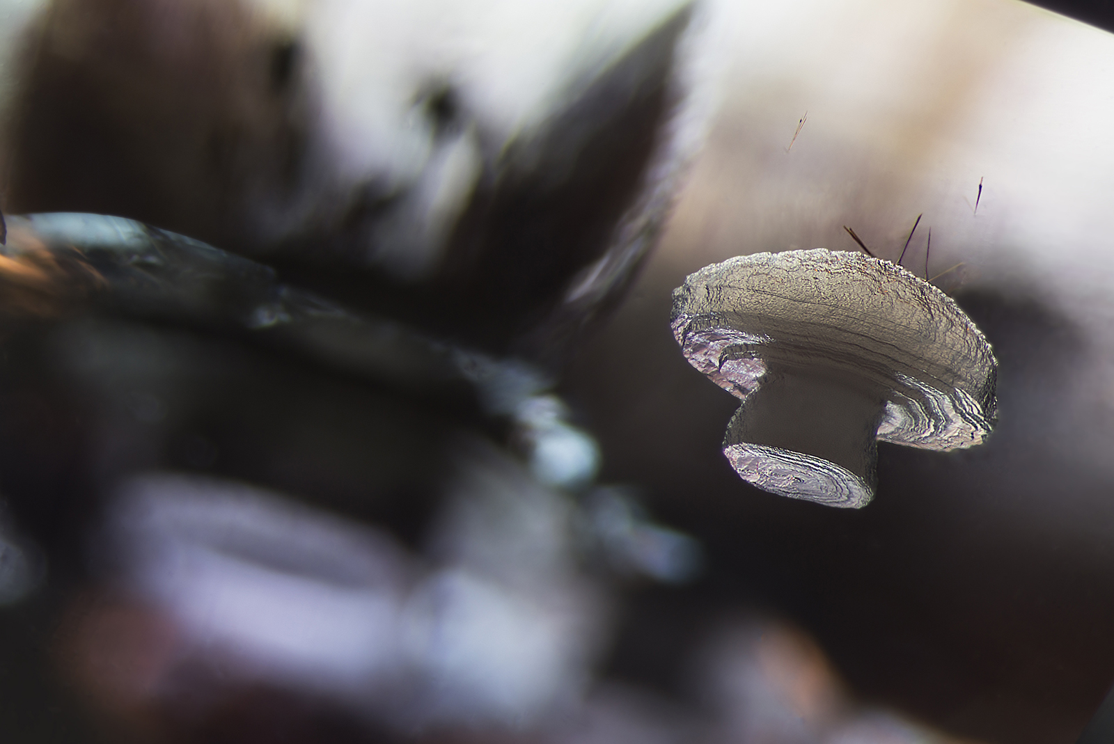
Lotus Gemology
Bangkok, Thailand
Cristobalite crystal suspended in its quartz mineral host
Darkfield
40x (Objective Lens Magnification)
19th Place – Martyna Lukoseviciute & Dr. Carrie Albertin
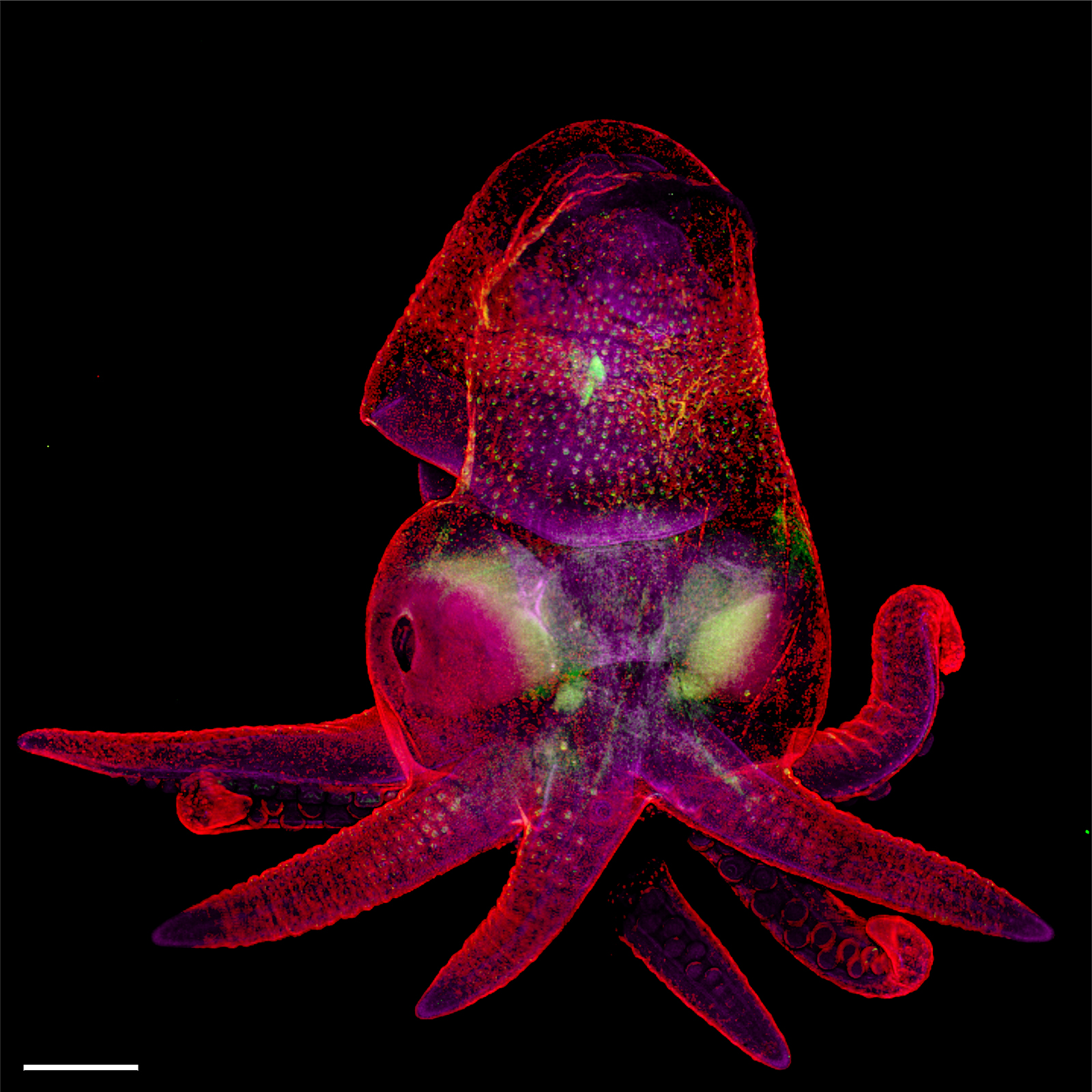
University of Oxford
Weatherall Institute of Molecular Medicine
Oxford, Oxfordshire, United Kingdom
Octopus bimaculoides embryo
Confocal, Image Stitching
5x (Objective Lens Magnification)
20th Place – Simon Merz, Lea Bornemann & Sebastian Korste
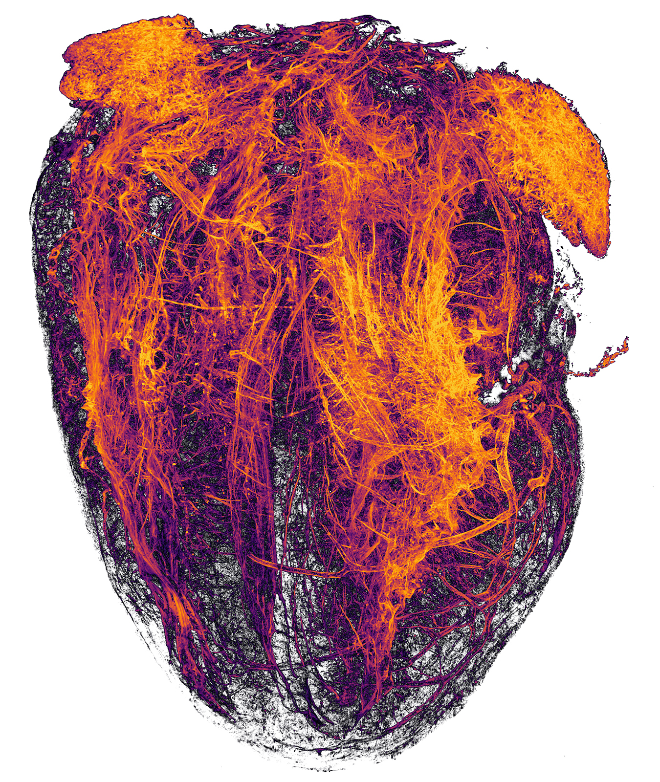
University Hospital Essen
Institute for Experimental Immunology & Imaging
Essen, Nordrhein-Westfalen, Germany
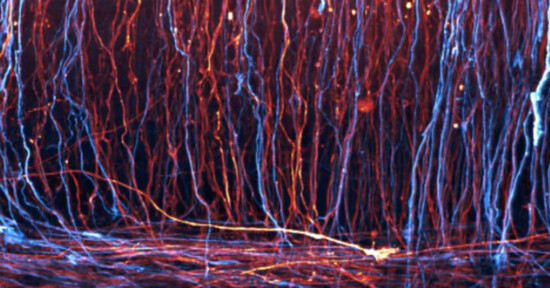
Blood vessels of a murine (mouse) heart following myocardial infarction (heart attack)
Tissue Clearing, Light Sheet Fluorescence Microscopy
2x (Objective Lens Magnification)
To see more of the incredible images that were honored by the Nikon Small World competition this year, or to learn more about this contest that sits at the intersection of art and science, head over to the Nikon Small World website.
Image credits: All images credited individually, shared courtesy of Nikon.