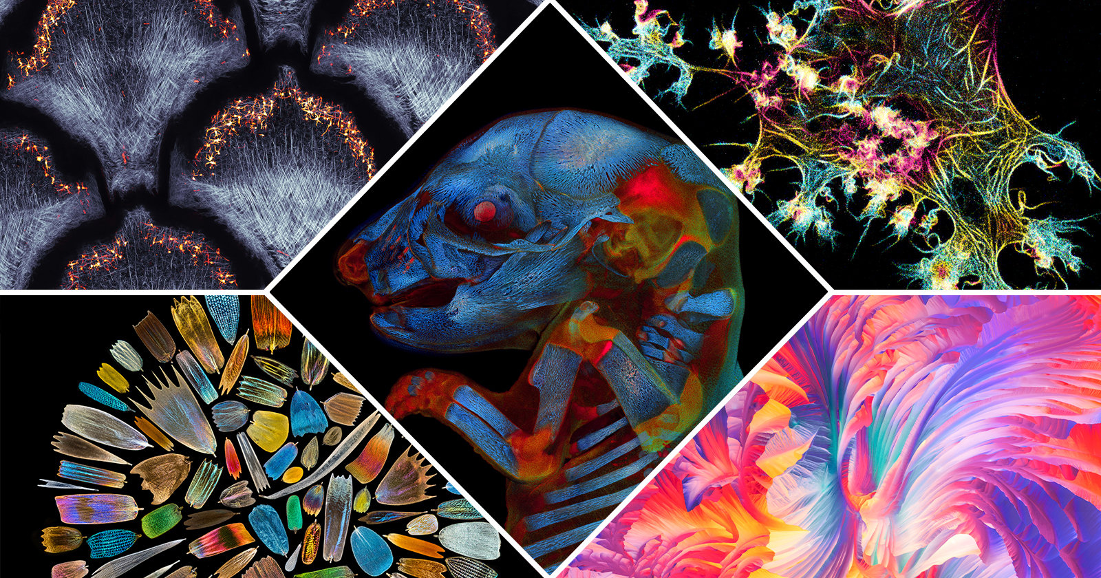
The Winners of the 2022 Nikon Small World Competition
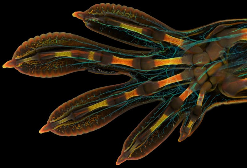
The winners of the 2022 Nikon Small World Photo Microscopy Competition have been announced, with a remarkably detailed photo of an embryonic Gecko hand taking the top prize.
Grigorii Timin and Dr. Michel Milinkovitch captured the winning image in the annual microscope photography competition with their image of a Madagascar giant day gecko’s front paw during development.
As part of his research under the supervision of Milinkovitch, Timin studied this embryo of the Phelsuma Grandis day gecko under the microscope. He used different fluorescent labels to stain distinctive parts of the gecko’s hand and the final result gives a glimpse into the hidden complexity of the creature.
Timin highlights the nerves of the gecko in a cyan color while the bones, tendons, ligaments, skin, and blood cells are stained in a range of warmer colors.
“This embryonic hand is about 3mm (0.12 in) in length, which is a huge sample for high-resolution microscopy,” says Timin. “The scan consists of 300 tiles, each containing about 250 optical sections, resulting in more than two days of acquisition and approximately 200GB of data.”
“This particular image is beautiful and informative, as an overview and also when you magnify it in a certain region, shedding light on how the structures are organized on a cellular level,” explains Timin.
He adds “The Nikon Small World Competition is a great opportunity to share how impressive nature is on a microscopic level, not only within a scientific community but also with the general public.”
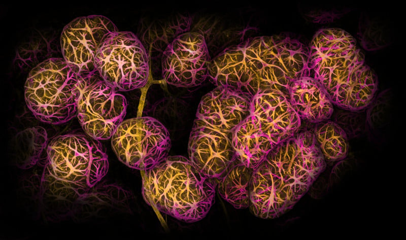
Second place was awarded to Dr. Caleb Dawson for his image of breast tissue showing contractile myoepithelial cells wrapped around milk-producing alveoli.
Taking a week to process, Dawson stained the myoepithelial cells with multiple rounds of fluorescent dyes and captured the image with a confocal microscope. Such images of lactating breast tissue can help researchers figure out how immune cells keep breast tissue and the babies it can feed healthy.
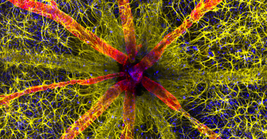
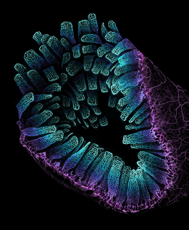
Third place was awarded to Satu Paavonsalo and Dr. Sinem Karaman for their image of blood vessel networks in the intestine of an adult mouse.
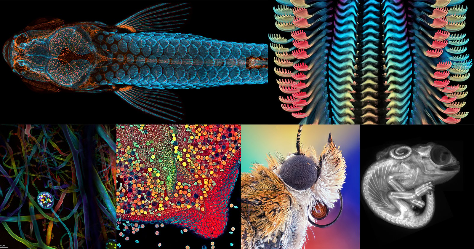
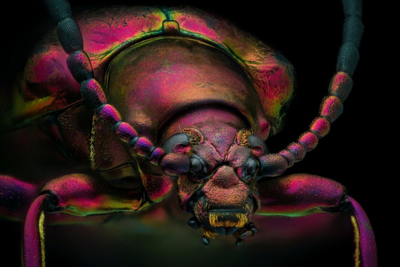
In addition to the top three winners, Nikon Small World recognized 89 photos out of thousands of entries from scientists and artists across the globe. Other highlights in the annual competition include a burning candle close-up, moth eggs, and a fly being savagely crushed by a tiger beetle.

More than 1,300 entries were received from 72 countries in 2022, the 48th year of the annual microscope photography competition. Modern microscopes are effectively very specialized camera systems with a powerful zoom, so it is not surprising that some of the images scientists take while they work are stunning artworks in their own right. However, the contest invites both photographers and scientists to submit images of all things visible under a microscope.
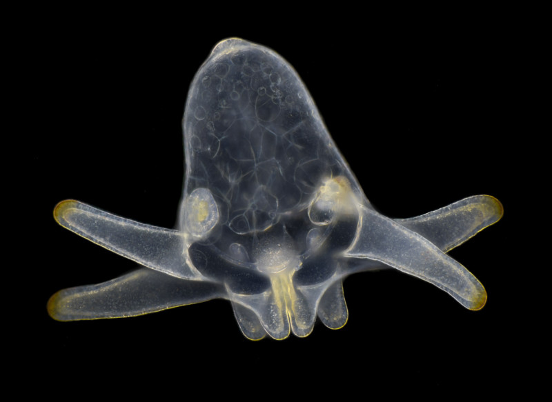
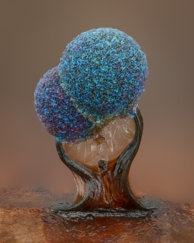
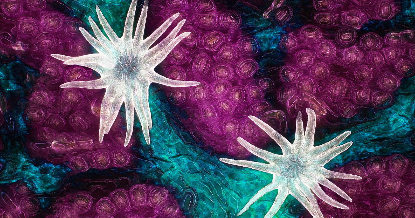
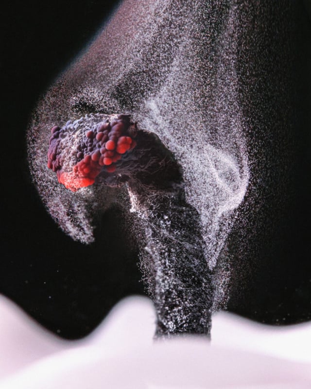
Wim van Egmond, a professionally trained photographer who specializes in microscope photography, received an honorable mention for his image of an anemone larva. Meanwhile, California photographer Allison Pollack specializes in mushroom photography and her extreme close-up of slime mold earned her fifth place in the competition this year.
“Each year, Nikon Small World receives an array of microscopic images that exhibit exemplary scientific technique and artistry. This year was no exception,” says Eric Flem, Communications and CRM Manager, at Nikon Instruments.
“At the intersection of art and science, this year’s competition highlights stunning imagery from scientists, artists, and photomicrographers of all experience levels and backgrounds from across the globe.”