Groundbreaking Close-Up of Brain Tumor Cells Wins Landmark 50th Nikon Small World Contest
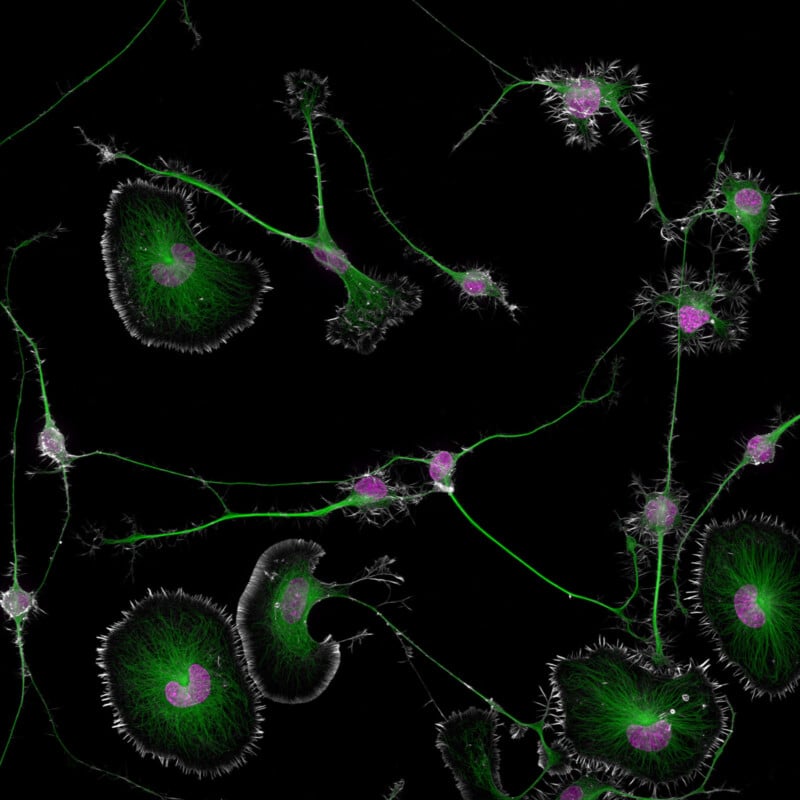
Nikon Instruments Inc. announced the winners of its 50th anniversary Nikon Small World Photomicrography Competition. This year, the prestigious photomicrography competition received around 2,100 entries from amateurs, researchers, students, and other scientists in 80 countries.
Founded in 1974, the Nikon Small World contest is widely considered the leading platform for recognizing the art, proficiency, technical skill, and photographic excellent involved in photomicrography, which is capturing images through a microscope. The Nikon Small World 2024 competition is joined by the Nikon Small World in Motion contest for video micrography, which unveiled its winners last month.
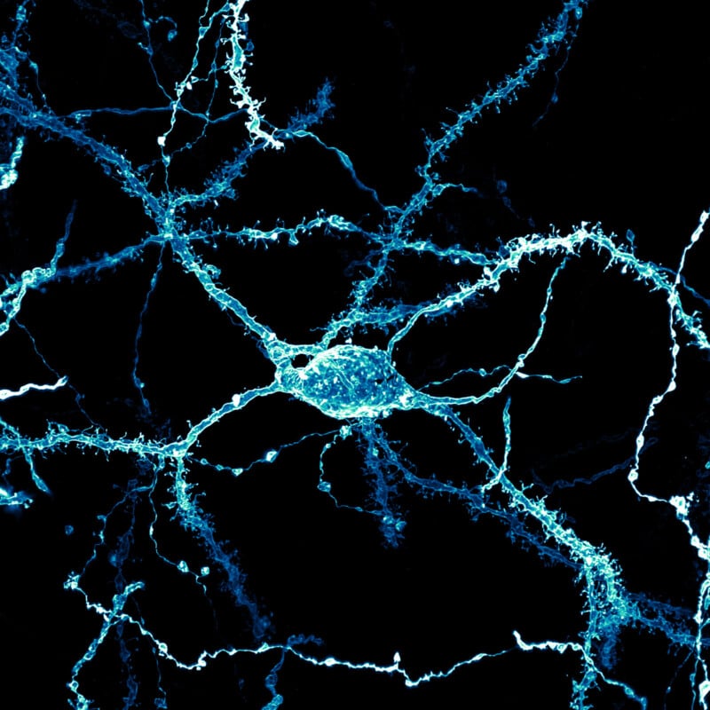
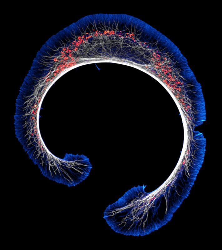
Dr. Bruno Cisterna, with assistance from Dr. Eric Vitriol of Augusta University, is this year’s winner for his remarkable image of differentiated mouse brain tumor cells showcasing the actin cytoskeleton, microtubules, and nuclei. This image shows how disruptions in the cell’s cytoskeleton, the structural framework of the cell, can lead to debilitating neurological diseases like Alzheimer’s and ALS.
Dr. Cisterna’s research is not just visually stunning; it revealed that the protein profilin 1 (PFN1), which is vital to building the cell’s structure, also plays a role in maintaining the microtubules — like little cellular highways — that are important for cellular transport.
“When PFN1 or related processes are disrupted, these highways can malfunction, leading to cellular damage similar to what is observed in neurodegenerative diseases,” Nikon Instruments explains.
“One of the main problems with neurodegenerative diseases is that we don’t fully understand what causes them,” says Dr. Cisterna. “To develop effective treatments, we need to figure out the basics first. Our research is crucial for uncovering this knowledge and ultimately finding a cure. Differentiated cells could be used to study how mutations or toxic proteins that cause Alzheimer’s or ALS alter neuronal morphology, as well as to screen potential drugs or gene therapies aimed at protecting neurons or restoring their function.”
Dr. Cisterna’s patience and skill were desperately needed to capture this photo, as he spent about three months “perfecting the staining process to ensure clear visibility of the cells.”
“After allowing five days for the cells to differentiate, I had to find the right field of view where the differentiated and non-differentiated cells interacted. This took about three hours of precise observation under the microscope to capture the right moment, involving many attempts and countless hours of work to get it just right,” Dr. Cisterna adds.
This hard work has not only paid off in the fantastic honor of winning the 50th Nikon Small World Photomicrography Contest, but Dr. Cisterna and his team’s research published four months ago in the Journal of Cell Biology may lead to a breakthrough in neurological research and help the millions of people battling neurodegenerative diseases. Research like this can transform lives in remarkable ways.
This spirit of scientific progress is reflected across many of the images entered into this year’s competition and the prior 49 editions.
“At 50 years, Nikon Small World is more than just an imaging competition — it’s become a gallery that pays tribute to the extraordinary individuals who make it possible. They are the driving force behind this event, masterfully blending science and art to reveal the wonders of the microscopic world and what we can learn from it to the public,” says Eric Flem, Senior Manager of CRM and Communications at Nikon Instruments.
“Sometimes, we overlook the tiny details of the world around us. Nikon Small World serves as a reminder to pause, appreciate the power and beauty of the little things, and to cultivate a deeper curiosity to explore and question,” Flem adds.
Second and Third Place
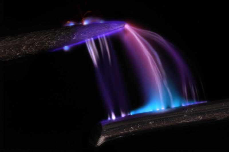
Dr. Marcel Clemens took home second place for an electrifying photo of electricity arcing between a pin and a wire.
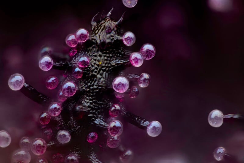
Chris Romaine earned the bronze for his image of a cannabis plant leaf. This image shows bulbous structures, trichomes, and cannabinoid vesicles, which are blister-like structures.
The Rest of the Top 20
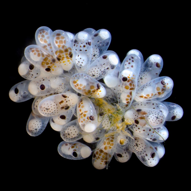
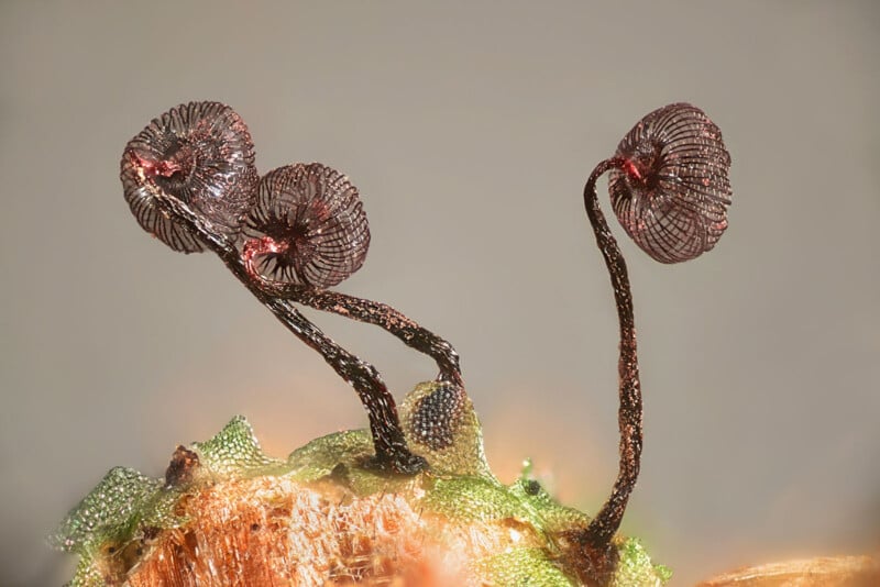
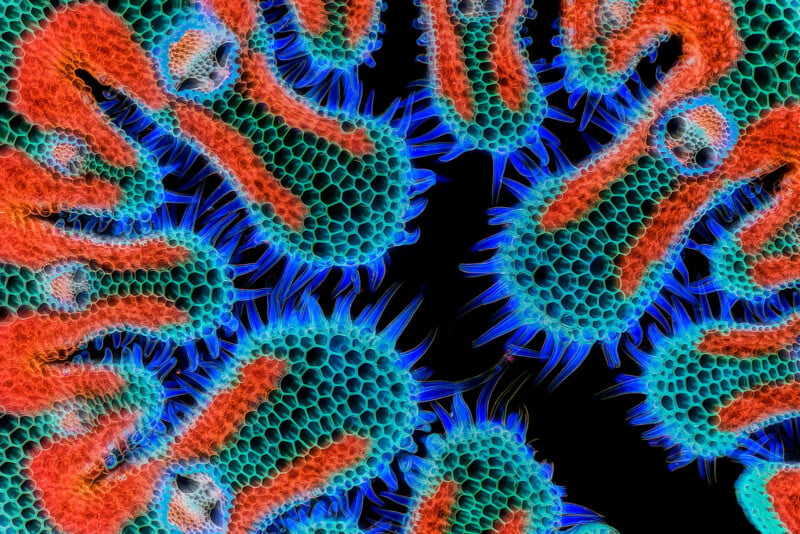
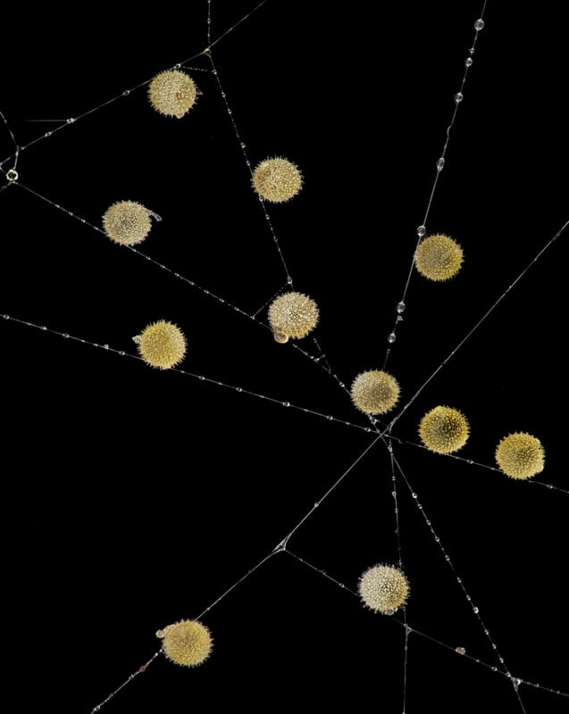
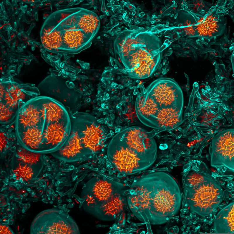
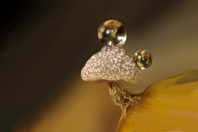
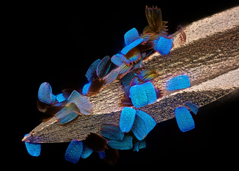
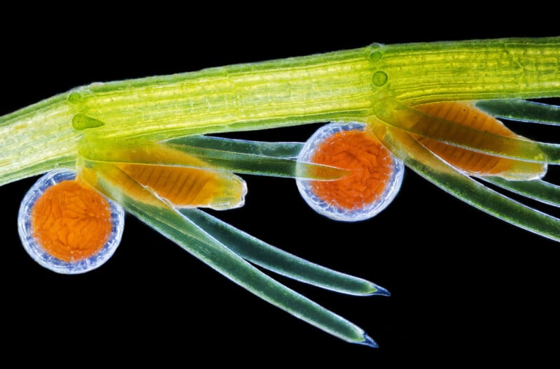
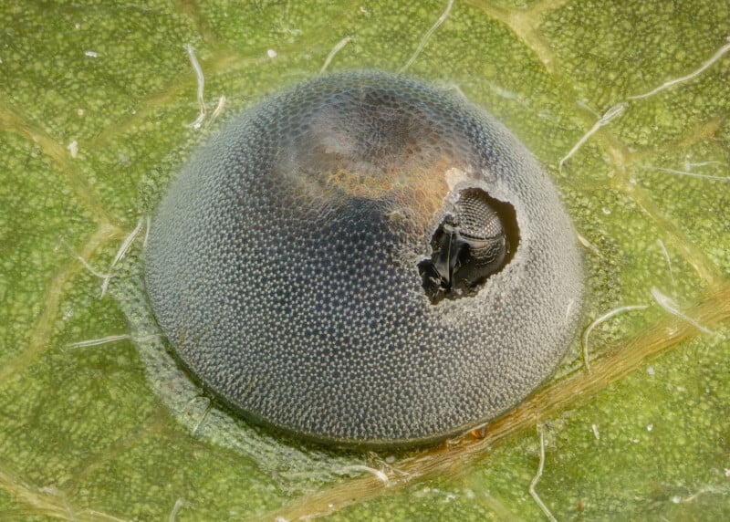
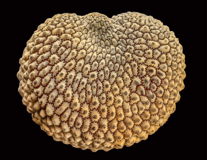
Alongside the top three winners, the Nikon Small World competition recognized 87 total images, all of which can be seen on the Nikon Small World Photomicrography Contest website. Below are the 17 other images comprising the top 20 for the Nikon Small World 2024 contest, presented in order from fourth to 20th place.
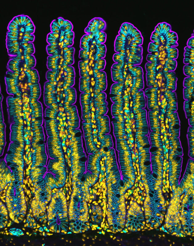
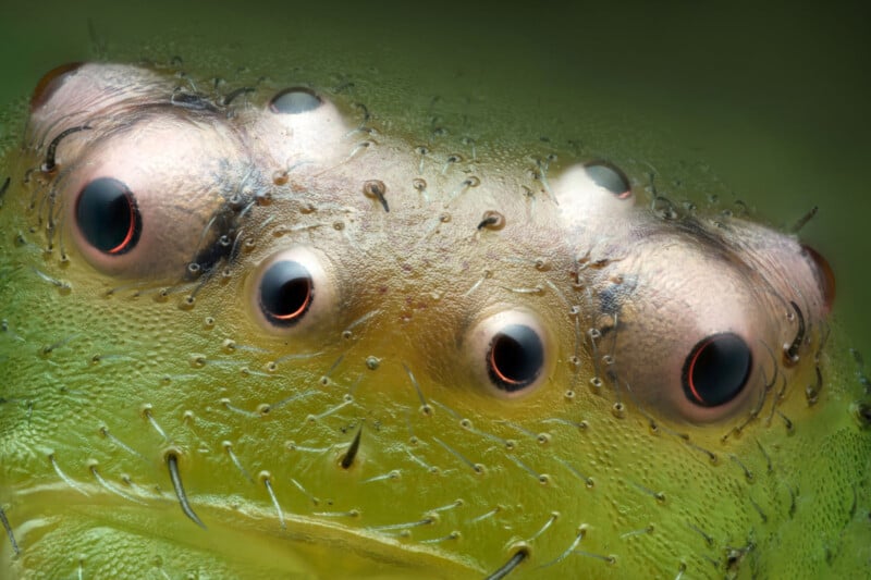
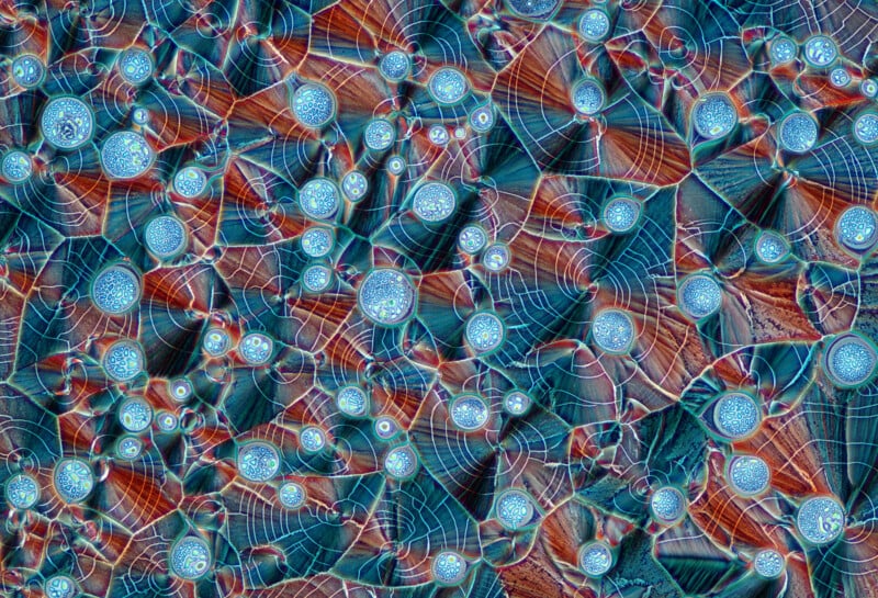
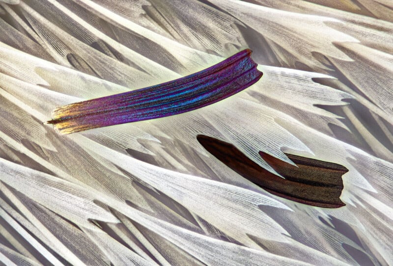
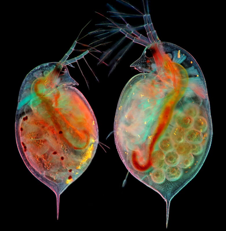
All recognized entrants can be viewed on the Nikon Small World website. It’s a remarkable competition, and there are many fascinating, beautiful microscope images to see. Even more photomicrographic goodness is available in PetaPixel‘s coverage of last year’s winners.
Image credits: All photos are individually credited and provided courtesy of Nikon Small World.