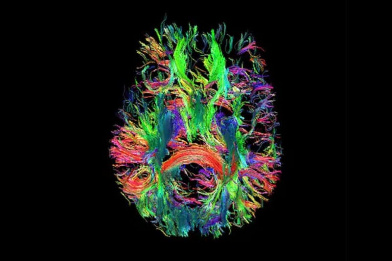
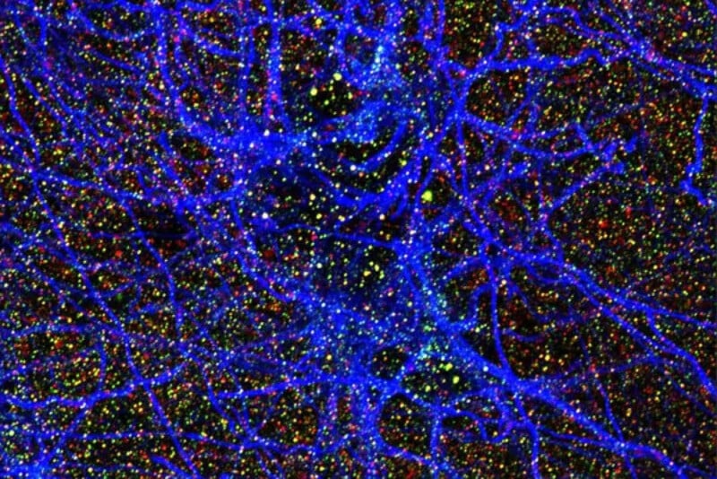
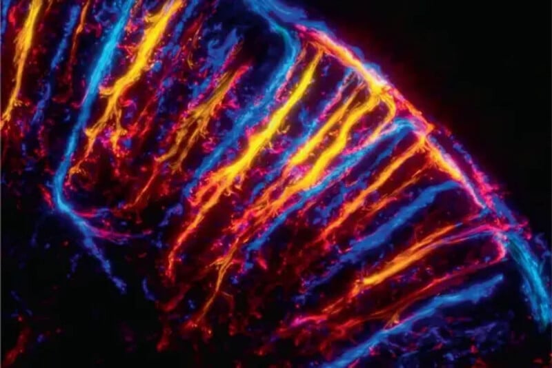
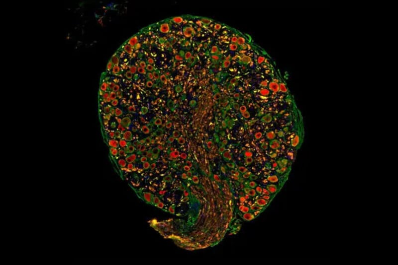
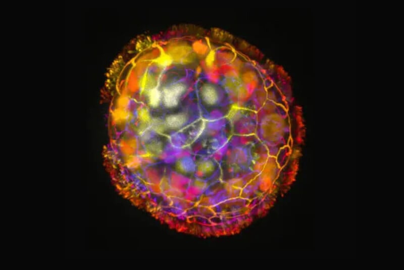
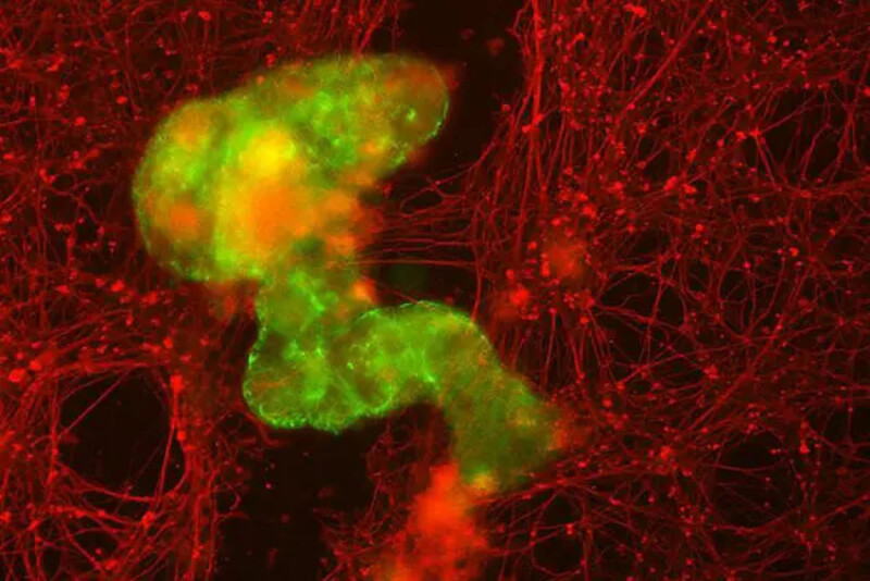
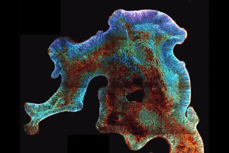
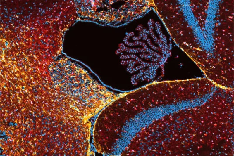
Mind-Blowing Images From the Field of Neuroscience

Neuroscience images

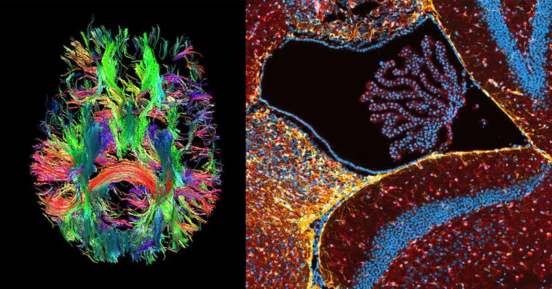
Although these images look as though they were created by an artist, they were actually taken by scientists trying to learn more about the human nervous system.

These stunning images were curated by The New York Times and are some of the very latest works from the field of neuroscience — the study of the nervous system with a primary focus on the brain.

A new MRI scanner captured this image which shows the complex circuitry of the human brain. The MRI scanner was developed at the University of California, Berkeley, and allows for 10 times higher resolution than its predecessors. Researchers hope they will be able to understand the processes behind the underlying illnesses like schizophrenia and autism. | An T. Vu, UCSF; David Feinberg and Alex Beckett, UC Berkeley and Advanced MRI Technologies
neuroscience image
The fluorescent dots in this image represent cells attacking the brain of a person with Alzheimer’s disease. Called an astrocyte, the cell is supposed to protect brain connections but by attacking the brain synapses it can contribute to the deterioration of a person’s mind. | Charles Zachary Klein, Raquel Taddei, Teresa Gomez-Isla, Massachusetts General Hospital
neuroscience image
This colorful image details microscopic blood flow in a rat’s brain in a good example of how new imaging tools capture higher levels of detail. The author also posted a video relating to this image (see below). | Alexandre Dizeux

neuroscience image
This remarkable image shows the trunk of a spinal nerve with neurons highlighted in green, immune cells in yellow dots, and tumor cells shown in red and orange dots. It’s a close-up of the devastating disease neurofibromatosis Type 2. | Sara Veiga and Christine Chiasson-MacKenzie at the Stott and McClatchey Labs at Massachusetts General Hospital
neuroscience image
This is a minuscule robot called an “Antrhobot” that scientists claim can crawl neural tissue in lab dishes to heal wounded areas. The tiny robots can be as thin as a strand of hair. | Gizem Gumuskaya et al., Advanced Science, Tufts University
neuroscience image
This is also a robot but in this image the minuscule robots have come together to become a “superbot”. The red strands show the growth of neurons in an area where they had been removed by scientists. | Gizem Gumuskaya et al., Advanced Science, Tufts University
neuroscience image
This brain cortex was grown by scientists so they could understand how the cortex develops and how pediatric brain tumors might also develop. | Anna Pagliaro, Benedetta Artegiani, Delilah Hendriks, Princess Maxima Center
neuroscience image
This image taken from the brain of a mouse that scientists had implanted with breast cancer cells shows how a network of capillaries comes under attack which ultimately grows the tumor. | Sonu Subudhi and Sophie Steinbuch, Massachusetts General Hospital
Discussion