New Gigapixel 3D Microscope Camera Captures Life in Incredible Detail
A group of researchers has created a new type of camera microscope that is able to capture gigapixel-scale images of extremely small subjects, allowing for a detailed and unprecedented view of life.
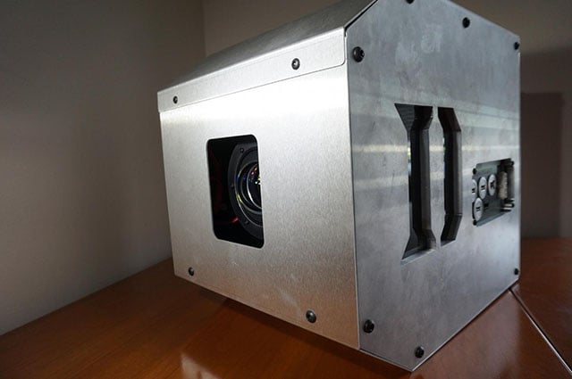
Developed by a group of scientists at the Computational Optics Lab at Duke University, the Multi-Camera Array Microscope (MCAM) is described as a large group of miniaturized digital microscopes that all acquire synchronized images and video in parallel. The team says they use a variety of algorithms to fuse the data acquired by the MCAM together to create a final gigapixel-scale composite.
“The easiest way to understand the MCAM’s capabilities is to digitally interact with some example image and video data that contains nearly one gigapixel (1 billion pixels) per frame,” the researchers explain.
So in addition to the video above that shows what is possible, further example image and video data are available for anyone to peruse on the group’s Gigaviewer webpage.
The idea was originally posed by a pair of Duke graduate students six years ago when they successfully combined 24 smartphone cameras into a single platform that was able to image an area about the size of a standard piece of paper. But over time and as they refined their system, they realized that they would be able to not only see incredible resolution at a sub-pixel level with their MCAM, but they would also be able to see the height of objects too.
“It’s like human vision,” Roarke Horstmeyer, assistant professor of biomedical engineering at Duke University, explains on the Pratt School of Engineering’s blog.
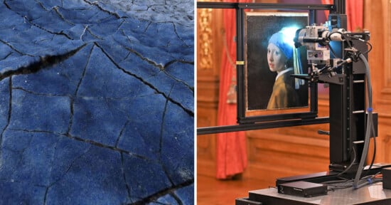
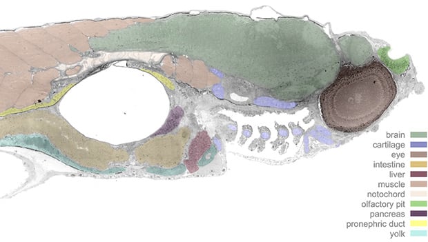
“If you merge multiple viewpoints together (as your two eyes do), you see objects from different angles, which gives you height. When our colleagues studying zebrafish used it for the first time, they were blown away. It immediately revealed new behaviors involving pitch and depth that they’d never seen before.”
The technology, which the group has three different designs for that offer different imaging specifications, is being developed further by the startup Ramona Optics, which is located right next to Duke University. The group has also published their research on gigapixel imaging at a microscopic level in a research paper.
“The modern laboratory is becoming more automated every day, with large well plates now being filled and maintained without ever touching a human hand,” Horstmeyer continues. “The sheer volume of data this is creating demands for new technologies that can help automate the tracking and capturing of the results.”
The group has shared multiple sample videos created by the MCAM as well as the raw files and open source code for those that are interested in learning more.