20 of the Most Beautiful Microscopic Photos in the World

smallworld_feat

If you thought it was impossible to take a stunning photo of cow dung, get prepared to be proven wrong. Cow dung, espresso coffee crystals, centipede fangs, these are just a few of the subjects that took the Top 20 spots in Nikon’s Small World competition.

The Nikon Small World competition is put on by Nikon Instruments, and it exists to shed a light on a genre of photos you don’t hear much about: photomicrography.

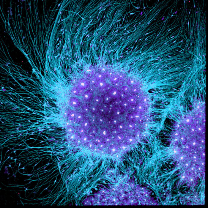
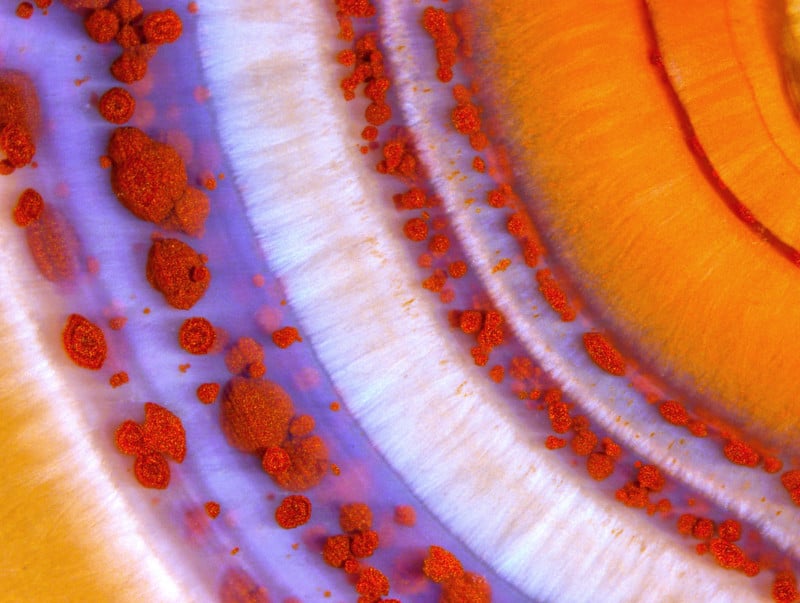
In layman’s terms, that’s photos taken with microscopes. And if you thought macro photography was cool, just wait until you get a load of these images. The top three prizes went to a photo (1) a photo of a three-day old zebrafish embryo, (2) a photo of a polished slab of Teepee Canyon agate, and (3) a culture of neurons derived from human skin cells.

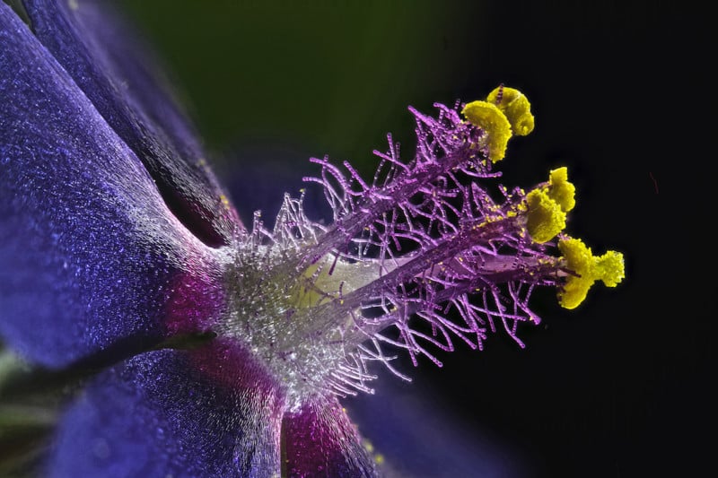
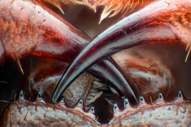
For our money, though, some of the photos that missed out on the top 3 spots are even more amazing. The wildflower stamens, espresso coffee crystals, and poison fangs of a centipede all blew us away. Check out all Top 20 (in order) below:
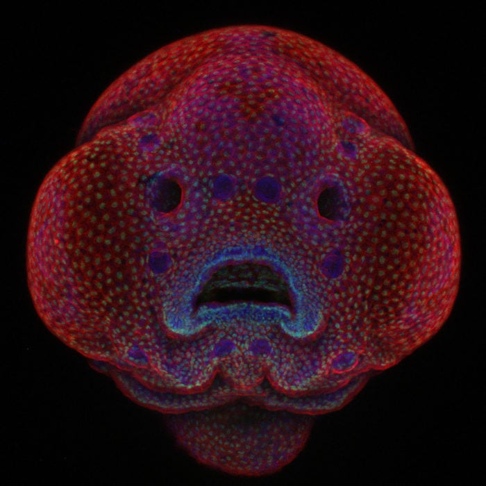
Four-day-old zebrafish embryo | Photo credit: Dr. Oscar Ruiz
Polished slab of Teepee Canyon agate | Photo credit: Douglas L. Moore
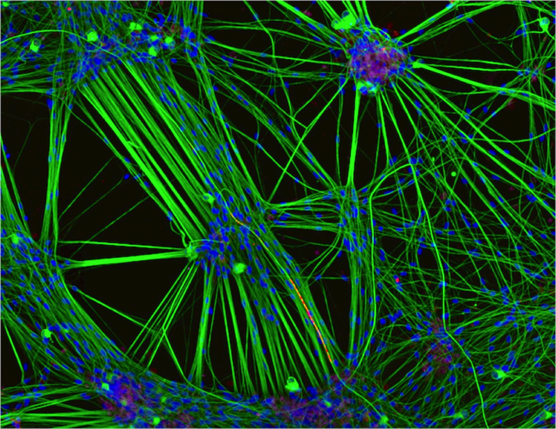
Culture of neurons (stained green) derived from human skin cells, and Schwann cells, a second type of brain cell (stained red) | Photo credit: Rebecca Nutbrown
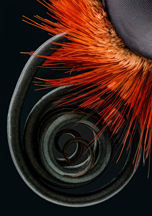
Butterfly proboscis | Photo credit: Jochen Schroeder
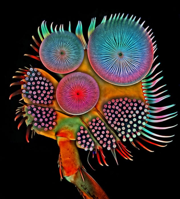
Front foot (tarsus) of a male diving beetle | Photo credit: Dr. Igor Siwanowicz
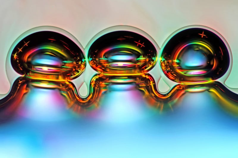
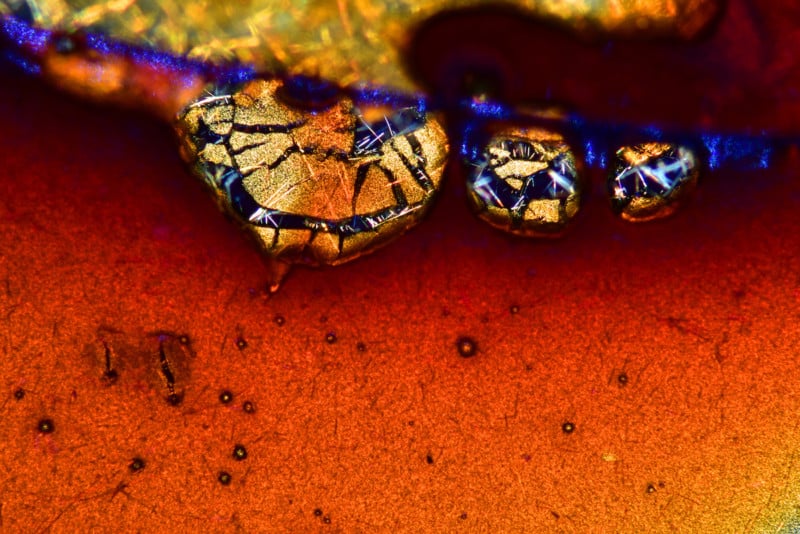
Air bubbles formed from melted ascorbic acid crystals | Photo credit: Marek Mis
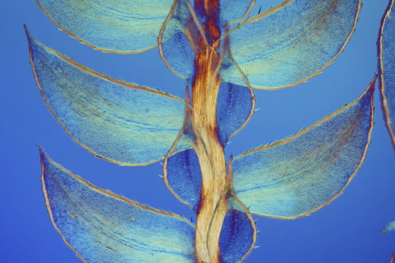
Leaves of Selaginella (lesser club moss) | Photo credit: Dr. David Maitland
Wildflower stamens | Photo credit: Samuel Silberman
Espresso coffee crystals | Photo credit: Vin Kitayama and Sanae Kitayama
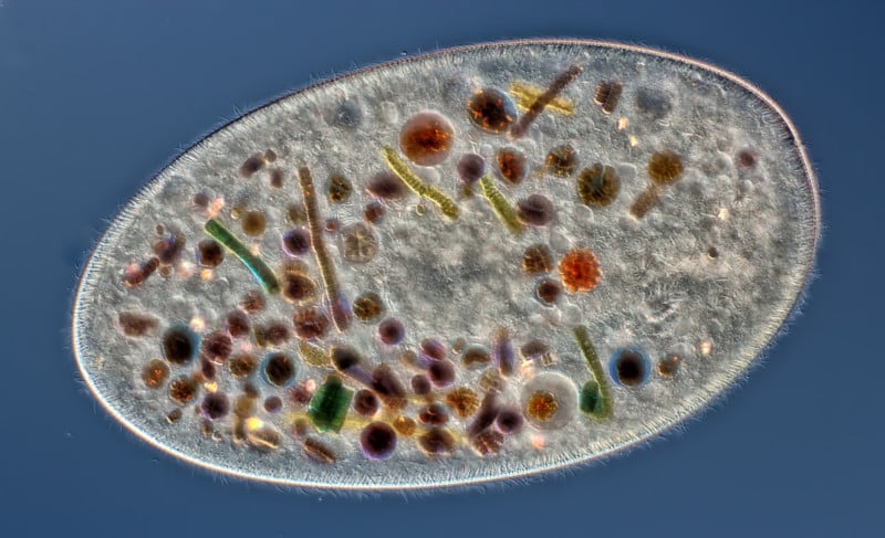
Frontonia (showing ingested food, cilia, mouth and trichocysts) | Photo credit: Rogelio Moreno Gill
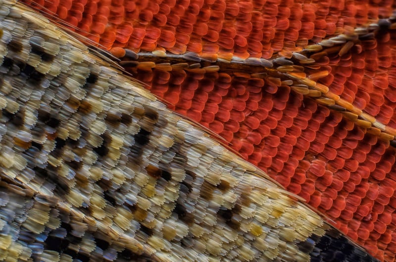
Scales of a butterfly wing underside (Vanessa atalanta) | Photo credit: Francis Sneyers
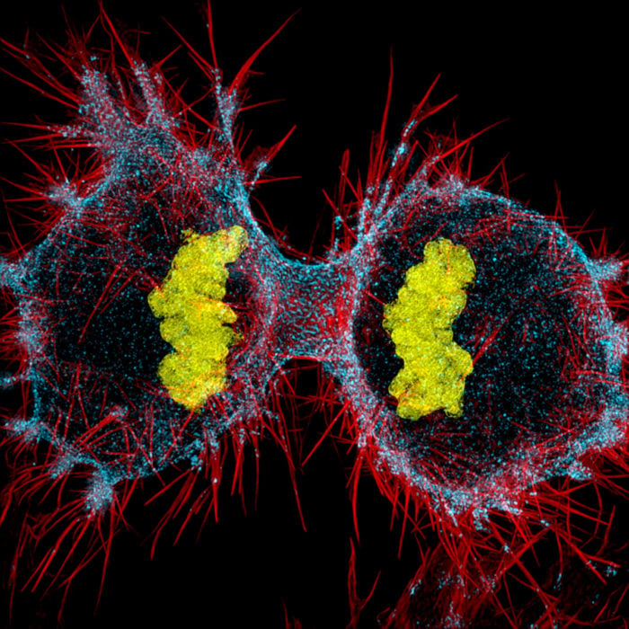
Human HeLa cell undergoing cell division | Photo credit: Dr. Dylan Burnette
Poison fangs of a centipede | Photo credit: Walter Piorkowski
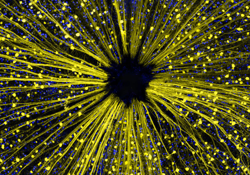
Mouse retinal ganglion cells  | Photo credit: Dr. Keunyoung Kim
Mouse retinal ganglion cells | Photo credit: Dr. Keunyoung Kim
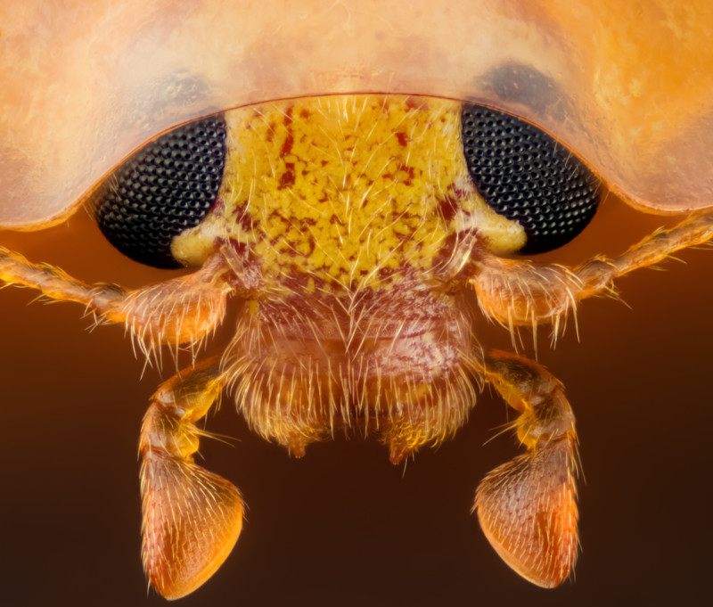
Head section of an orange ladybird | Photo credit: Geir Drange
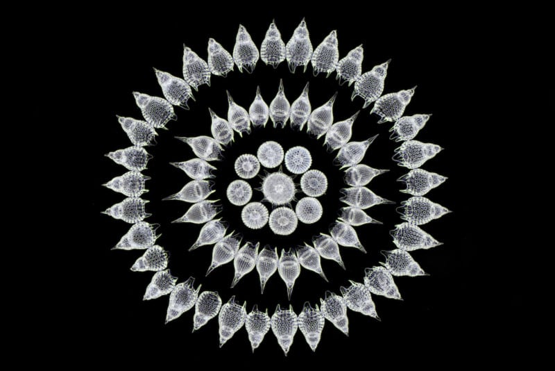
65 fossil Radiolarians (zooplankton) carefully arranged by hand in Victorian style | Photo credit: Stefano Barone
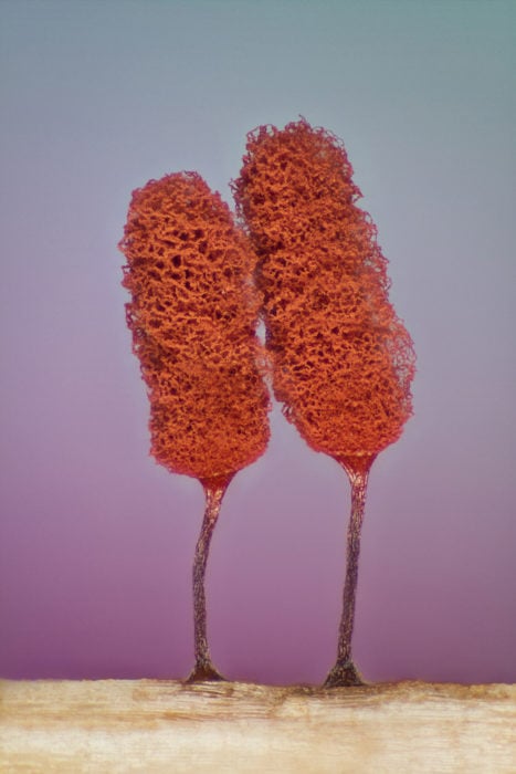
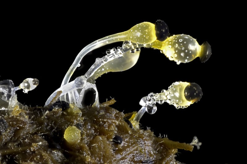
Slime mold (Mixomicete) | Photo credit: Jose Almodovar
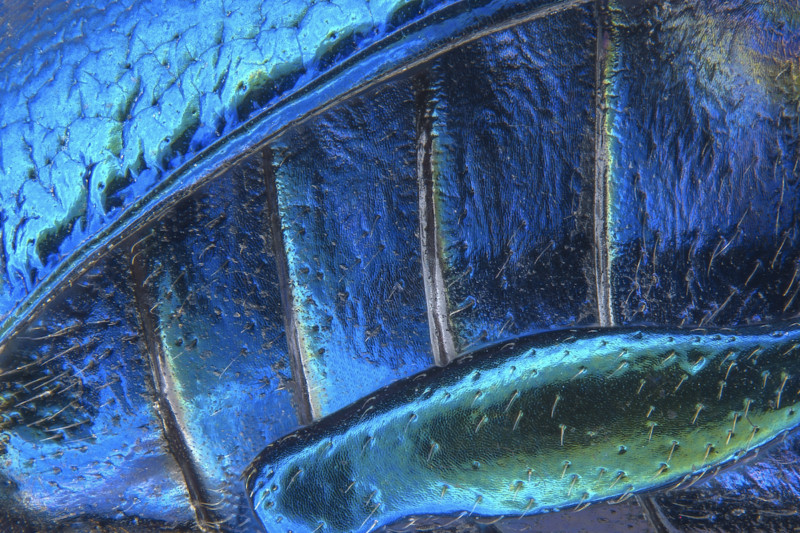
Parts of wing-cover (elytron), abdominal segments and hind leg of a broad-shouldered leaf beetle | Photo credit: Pia Scanlon
Human neural rosette primordial brain cells, differentiated from embryonic stem cells | Photo credit: Dr. Gist F. Croft, Lauren Pietilla, Stephanie Tse, Dr. Szilvia Galgoczi, Maria Fenner, Dr. Ali H. Brivanlou
Cow dung | Photo credit: Michael Crutchley

If you, like us, have some favorites that you think fell a bit too far down the list, it’s worth noting that Nikon is letting you vote for your favorite. Click here for the “Popular Vote” part of the competition and let the judges know which photo you think should have won.

And if you want to find out more about the the Nikon Small World competition, head over to their website or give the competition a follow on the newly-created Nikon Instruments Instagram account.


Image credits: All photos credited individually, used with permission from Nikon.

Discussion