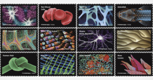
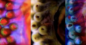
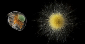
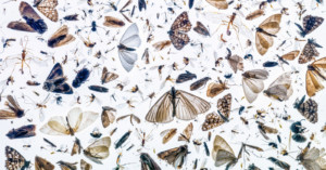
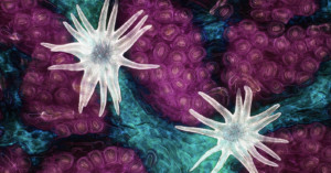
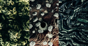
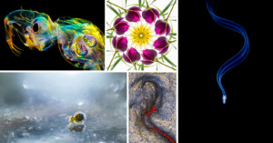
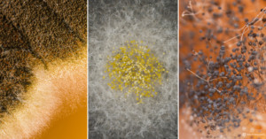
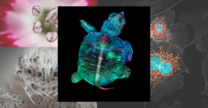
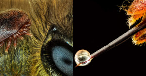
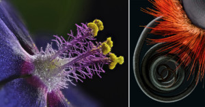
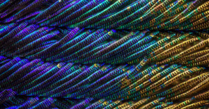
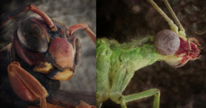
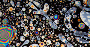
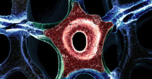
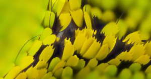
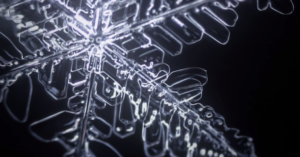
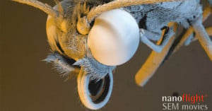
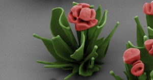
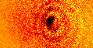
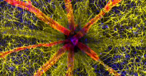
Harvard School of Engineering and Applied Sciences postdoctoral fellow Wim L. Noorduin, along with his colleagues, have discovered an interesting way to make pictures of flowers from microscopic crystals, as seen under an electron microscope.
Read more…

David Scharf is a basement pioneer in the art of making some of the world’s smallest things appear huge.
Read more…

Jack over at the astrophotography blog The Landingfield has published a series of photographs showing what a digital camera’s CMOS sensor looks like when viewed through a microscope. The sensor (seen above) was taken from a broken Nikon D2H — a DSLR from back in the early 2000s.
Read more…

This image might look like some kind of screenshot from an old 16-bit video game, but it’s actually the first photo ever made of an atom’s shadow. Researchers at Griffith University in Australia suspended a ytterbium atom in midair, shot it with a laser beam, and then used a Fresnel lens on the other side to snap a photograph of the dark shadow left by the atom. Scientist Erik Streed has a writeup explaining how they accomplished it and the project’s implications for other research.
Snapping an atom’s shadow? Now that’s a first (via Engadget)
Image credits: Photograph by Kielpinksi Group/Centre for Quantum Dynamics

A team of researchers at the Japan Science and Technology Agency have recently taken 3D imaging to another level. A much smaller level. What they’ve managed to do is develop an electron microscope that can show 3-dimensional photos of their tiny subjects in real-time. In the past, getting a 3D photo from an electron microscope meant superimposing two images taken at slightly different angles. But this new microscope allows the scientist to slant the electron beam and obtain both angles at the same time.
The resulting images are slightly lower in resolution, but the advantage of seeing their structure in 3 dimensions makes it a worthwhile tradeoff. And all this without the need for 3D glasses.
(via Gizmodo via The Verge)
Photographer Clemens Wirth wanted to dive into microscopy, so he attached his Canon 5D Mark II to a monocular microscope using an adapter and pointed it at one small drop of water. He was amazed to find out how much activity goes on inside ordinary water, and how detailed that tiny world is. This short film, titled “Micro Empire”, is a beautiful combination of Wirth’s footage and audio by Radium Audio.
(via Laughing Squid)

A team of researchers at UC Davis have come up with a super-cheap way of turning an iPhone into a microscope — useful for diagnosing diseases in areas where medical equipment is hard to come by. Inspired by the CellScope project at UC Berkeley, Sebastian Wachsmann-Hogiu decided to create something even smaller and cheaper. By taping a 1-millimeter ball lens embedded in a rubber sheet to the iPhone, he was able to boost magnification by 5x, which allows the camera to photograph blood cells. Only a small portion of each image is in focus, so they also utilize focus stacking to achieve more usable photos.
The best part is the price — each lens only costs $30-40, and would be even cheaper if mass produced.
The Research Paper (via Digital Trends)

Having a camera that shoots 5000 frames per second is enough to capture slow motion footage of a bullet flying through the air, but scientists at the Science and Technology Facilities Council have now announced a camera that shoots a staggering 4.5 million frames per second. Rather than bullets, the camera is designed to capture 3D images of individual molecules using powerful x-ray flashes that last one hundred million billionth of a second. The £3 million camera will land in scientists hands in 2015.
(via STFC via Engadget)
P.S. In case you’re wondering, the image above is an illustration showing x-ray flashes being generated.

Thought the grain-of-salt-sized camera announced in Germany earlier this year was small? Well, researchers at Cornell have created a camera just 1/100th of a millimeter thick and 1mm on each size that has no lens or moving parts. The Planar Fourier Capture Array (PFCA) is simply a flat piece of doped silicon that cost just a few cents each. After light information is gathered, some fancy mathematical magic (i.e. the Fourier transform) turns the information into a 20×20 pixel “photo”. The fuzzy photo of the Mona Lisa above was shot using this camera.
Obviously, the camera won’t be very useful for ordinary photography, but it could potentially be extremely useful in science, medicine, and gadgets.
(via Cornell Chronicle via Engadget)

If you had a camera the size of a grain of rice, that would be considered extremely small, but researchers at the Fraunhofer Institute in Germany have created a camera the size of a grain of salt. The world’s smallest camera measures 1x1x1 millimeters, shoots 0.1 megapixel photographs (250×250 pixels), and is so inexpensive to make that they’re disposable. Potential uses for the camera include photographing the inside of human bodies (AKA endoscopy) and being used as rearview cameras on cars.
Cameras out of the salt shaker (via CrunchGear)