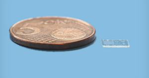
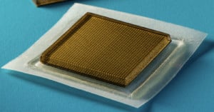
An interesting new imaging technique in use by fertility experts at the CARE fertility clinic in the United Kingdom may be the key to increasing the likelihood of a successful a IVF therapy. The process involves snapping thousands of images of embryos in development in order to help doctors better select which embryos to implant successfully.
Read more…

Most photographers and artists will never have the opportunity to make the kind of images that Andy Ellison does. As an MRI technologist at Boston University Medical School, Ellison has access to extremely expensive imaging machines. More specifically, he runs a research-only Philips 3 Tesla MRI machine. When he’s not using it for official purposes, he experiments with it by placing various fruits, vegetables, and flowers inside. The resulting still images and animations are beautiful and abstract, and form a project that he calls “Inside Insides.” The images above show a pineapple and an artichoke.
Read more…

There’s an interesting discussion going on over at the DPReview forums regarding how the human eye compares to the technology we have in digital cameras.
Here are some of the findings that were compiled from various sources on the web:
- Sensor size: 22mm in diameter
- Resolution: 576 megapixels
- Sensitivity: 1 – 800 ISO
- Focal length: 22mm – 35mm
- Aperture: f/2.1 – f/8.3
Another interesting idea that came up was the possibility of using the human eye as the lens and sensor for future imaging devices:
Maybe future “cameras” will actually link to your eyes – since the eyeball is such a great lens, who knows? Getting signal from the eye is the trick – would require a surgical implant or a means of reading brainwaves. Maybe that’s 200 years out – similar time [frame] the Mayo clinic is talking about for correcting double/triple vision.
Perhaps in the future we’ll all be documenting our lives at 576 megapixels through our eyes and ears, and storing the photos and videos on petabyte external hard drives at home.
What do you think of this discussion? Is there anything that jumps out at you as being wrong, or do you agree with the comparison for the most part?
Image credit: Eye (please add some funny TAGs) by Michele Catania
Ever wonder what a vinyl record looks like under an electron microscope? Okay, probably not. Luckily, there’s people who do, including Chris Supranowitz, who created a number of electron microscope images for a course at the University of Rochester.
Here’s a photograph of the record grooves captured by Supranowitz at 500x magnification. Those dark chunks you see are dust particles.

This one was shot at 1000x magnification. The record begins to look like the Grand Canyon.

These images were created in the Spring of 2005 for the course Opt 307/407: Practical Electron Microscopy and Advanced Topics. Other projects used the electron microscope to examine such things as snowflakes and bird feathers.
To see more of the amazing images captured by Supranowitz, check out the final project page.
(via Reckon)